Imaging Shows how COVID-19 Affects the Brain

COVID-19 affects the lungs to make it hard to breath, but it can also affect other areas of the body, such as the heart and kidneys. A number of recent radiology studies using computed tomography (CT) and magnetic resonance imaging (MRI) show that infection with coronavirus, which causes COVID-19, can also affect the brain and nervous system.
Doctors have known for years that viral infections can cause systemic effects, or effects occurring throughout multiple organs and systems throughout the body, far away from the original source of infection. Now they are discovering that COVID-19 can have systemic effects – it can even cause neurological problems, such as dizziness, difficulty concentrating, a loss of smell and taste, seizures, strokes, and weakness. In fact, research shows that about half of all patients with COVID-19 experience neurological symptoms.
Viral infections can cause serious systemic effects and neurological problems, such as:
Encephalopathy – a general term for any brain disease that alters the structure or function of the brain
Critical illness myopathy – a type of muscle disease that can develop in intensive care unit (ICU) patients who receive steroids or other neuromuscular drugs
Neuropathy – damage or weakness to one or more nerves, resulting in numbness, tingling, muscle weakness and pain in the affected area
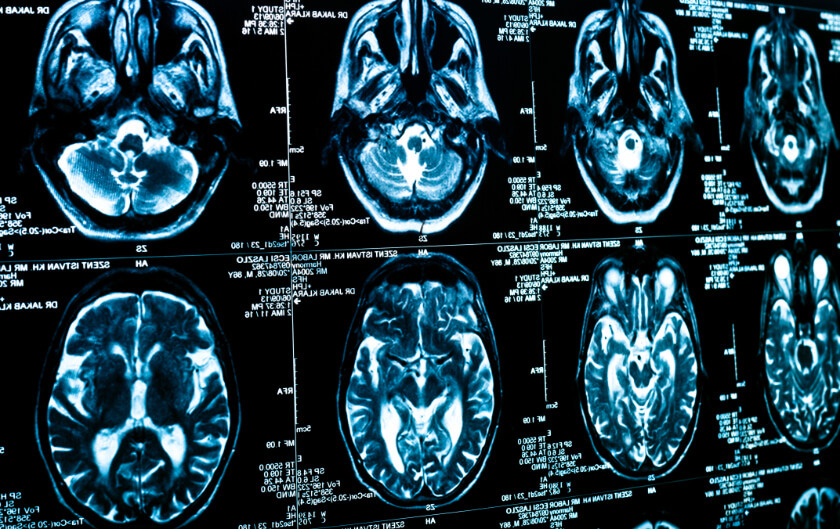
Infections like COVID-19 can also damage the brain’s white matter, which is the tissue that helps pass messages through the brain’s grey matter, and even cause lesions and bleeding there. Doctors can use MRI to detect bleeding and other damage to the white matter. In a study of 11 critically ill COVID-19 patients with persistently depressed mental status, doctors found evidence of lesions in 10 of the 11 patients, and bleeding in seven.
Coronavirus infections can damage the nervous system in a number of ways. Scientists can detect the virus’s proteins and genetic material in fluid and tissues of the spine and brain, which suggests viruses could invade the nervous system directly to cause nerve damage. Coronavirus may cause neurological damage by triggering an excessive immune response, known as a cytokine storm. Cytokines can cross the blood-brain barrier to cause a type of brain damage known as acute necrotizing encephalopathy. Excessive immune response might also cause Guillain-Barré syndrome (GBS), a rare neurological disorder in which the immune system mistakenly attacks the peripheral nerves outside the brain and spine. Doctors can see evidence of some of these effects on CT and MRI images.
Researchers Use CT and MRI to Detect Neurological Damage Associated with COVID-19
Many patients with COVID-19 experience neurological symptoms, such as nausea, vomiting, headache and a loss of taste and smell. Others are suffering strokes, seizures, and other serious neurological effects. While these neurological symptoms often take a back seat to breathing problems in patients critically ill with COVID-19, they can have long lasting effects. In some cases, the neurological damage done by coronavirus can be life threatening.
In a study of 108 hospitalized COVID patients in Italy, for example, 59 percent of the participants had altered mental function and 31 percent had suffered a stroke. The researchers performed CT and MRI scans on the participants. These scans show that COVID affects the brain in serious ways.
The imaging tests showed that nearly one-third of the patients in the study had suffered strokes. Specifically, they suffered ischemic stroke, which occur when a blood clot blocks a blood vessel to the brain and prevents the brain from getting the oxygen-rich blood it needs.
Imaging revealed that six of the participants had suffered intracranial hemorrhage, or bleeding on the brain. Two had cerebral venous thrombosis, which is a condition in which a blood clot prevents blood from draining from the brain; the pressure inside the vein can cause blood to leak into brain tissue and potentially cause a hemorrhage or brain swelling.
The images obtained in the study showed that one patient in the study had developed a rare nerve disease, known as Miller-Fisher syndrome, which causes abnormal muscle coordination, paralysis of the eye muscles and absence of tendon reflexes, such as the classic “knee jerk” response. Viral illness, like that caused by coronavirus, typically precedes Miller-Fisher syndrome. Another participant’s radiological images showed signs of posterior reversible encephalopathy syndrome (PRES), a condition that causes headaches, seizures, altered mental status, and visual loss.
Because coronavirus infections can affect the nervous system, and because COVID-19 can cause immune responses that turn these infections into neurological diseases, many doctors now use MRI and CT imaging when evaluating COVID-19 patients for neurological symptoms. These images can help doctors make decisions regarding care for critically ill patients.
For more information on how imaging, such as CT and MRI scans can help doctors provide care for COVID-19 patients, contact your doctor or radiologist.
Related Posts

Why Do I Have to Fast Before Some Medical Imaging Tests?
When it comes to medical imaging, getting a clear, accurate image is a top priority. After all,

The Role of Radiology in the Coronavirus
Infections with the novel coronavirus began in Wuhan, China, at the end of 2019. Other countries
Cancer Deaths Increase Worldwide – Early Detection Can Save Your Life
Despite advances in medical technology to detect and treat cancer, a new study shows that the nu