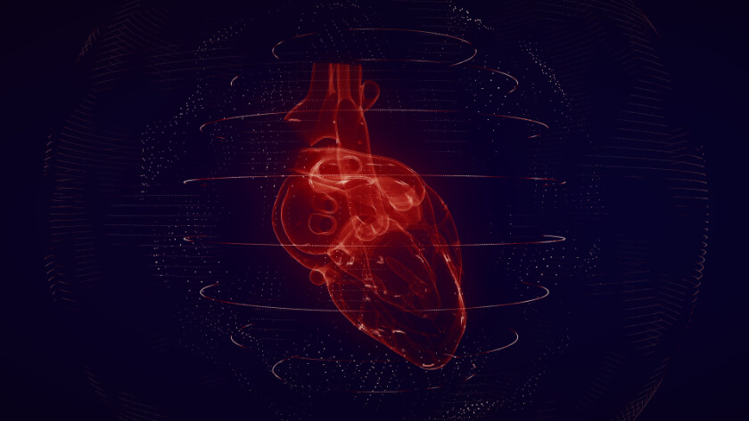
Using MRI to Identify Patients with Sarcoidosis at Risk for Bad Cardiac Outcomes

Some people with sarcoidosis experience adverse outcomes that cause severe illness and even death, while other people with the condition do not. Determining which patients with sarcoidosis are at higher risk for adverse outcomes can potentially save thousands of lives each year. The results of a new study suggest that MRIs may someday help doctors identify patients with sarcoidosis of the heart who are at risk for serious cardiac complications.
Sarcoidosis is a rare condition in which immune cells accumulate to cause clumps of tissue and inflammation that change how the affected tissues and organs work.
The clumps, known as granulomas, can develop anywhere in the body – including in the heart. The granulomas associated with cardiac sarcoidosis can cause inflammation, or swelling, which prevents the heart from pumping blood efficiently. The changes associated with cardiac sarcoidosis can cause serious heart problems, such as heart rhythm problems and even sudden cardiac arrest.
Sarcoidosis is a rare disease that affects about 200,000 Americans. Cardiac sarcoidosis is even more rare: doctors detect cardiac sarcoidosis in only about 2 to 5 percent of patients with systemic sarcoidosis, which is sarcoidosis in other parts of the body. Some postmortem reports suggest that the incidence may be as high as 20 to 30 percent.
Cardiac sarcoidosis causes a number of symptoms, including:
- Chest pain
- Fainting
- Fatigue
- Irregular heartbeats known as arrhythmias
- Palpitations, which are skipped or irregular heartbeats
- Shortness of breath
- Swelling in the legs from fluid excess
Treatment for cardiac sarcoidosis includes:
Implantable Cardioverter Defibrillator (ICD) – a small electronic device surgically implanted into the heart where it continuously monitors and helps regulate fast and potentially life-threatening electrical problems within the heart
Pacemaker – an electronic device implanted into the chest where it helps regulate slow electrical problems within the heart
Catheter ablation – doctors perform catheter ablation to treat arrhythmias; the procedure involves threading a long, flexible tube through blood vessels to the heart then transmitting heat or cold to destroy the faulty heart tissue cells causing the arrhythmia
Heart transplant/left ventricular assist device – replacement of a diseased heart with a healthy one
Currently, doctors use MRI to help diagnose people with sarcoidosis. In the results of a new study, researchers suggest that doctors may use MRI to predict which patients with sarcoidosis are likely to experience adverse events.
MRI can Help Doctors Diagnose Cardiac Sarcoidosis
Diagnosing cardiac sarcoidosis can be difficult because the condition causes very few signs and symptoms in its early stages. When symptoms do occur, they often mimic signs and symptoms of other disorders. Diagnosis requires thorough testing that may include electrocardiogram (EKG), echocardiogram (echo), and biopsy. Cardiac MRI (magnetic resonance imaging) is essential.
MRIs use a large magnet, radio waves, and a computer to make images of specific body organs. A cardiac MRI creates images of the heart. The large, tube-shaped MRI machine creates a strong magnetic field that lines up hydrogen protons in the body; the radio waves then knock the protons out of position. As they realign back into their proper positions, the protons send out radio waves that the computer receives and converts into images of the heart.
Cardiac sarcoidosis damages the heart in ways that are visible on MRI images. Radiologists use a technique known as late gadolinium enhancement (LGE) to look for evidence of cardiac sarcoidosis in the form of scar tissue. The presence of scar tissue on LGE means the patient may have cardiac sarcoidosis. The pattern and extent of the scar tissue may predict adverse events, such as heart failure and sudden death.
Healthcare professionals also use MRI to assess left ventricle ejection fraction (LVEF), which is the amount of blood the heart’s left ventricle pumps out with each heartbeat. Poor LVEF means the heart is not pumping blood efficiently enough to provide the body with the oxygen- and nutrient-rich blood it needs to function well.
MRI may also help doctors predict which patients will experience adverse outcome from cardiac sarcoidosis
According to the findings in a new study published in JAMA Cardiology, patients with certain features on MRI may be at higher risk for adverse outcomes from cardiac sarcoidosis. The new study builds on previous research that described the basic features of damage in the hearts of cardiac sarcoidosis patients who died suddenly or who needed a heart transplant.
Researchers enrolled 504 patients into the study, and categorized the participants according to their LVEF and LGE results on MRI. They found that patients with sarcoidosis who had large areas of scar tissue evident in LGE had a high rate of heart arrhythmias and heart failure events, even if their LVEF was normal.
The researchers encouraged other scientists to replicate the study in larger, more diverse groups of participants. They also suggested further study into whether the features detected on MRIs should guide the treatment in patients with cardiac sarcoidosis.
Related Posts
Chest X-rays Can Help Doctors Predict the Severity of COVID-19 in Young and Middle-Age ER Patients
Like many diseases, COVID-19 can be unpredictable. For example, the coronavirus that causes COVI

3D Mammography Provides Special Benefits for Older Women
Mammography is an effective breast cancer screening method for women ages 65 and older, but the

How Smoking Causes Lung Cancer
Doctors have known for years that smoking cigarettes, cigars, and pipe tobacco causes cancer. In