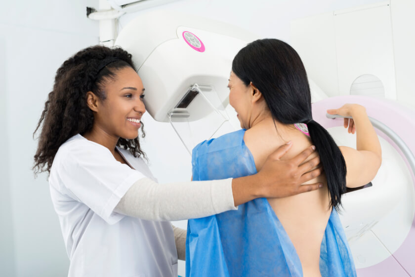
What is 3D mammography?
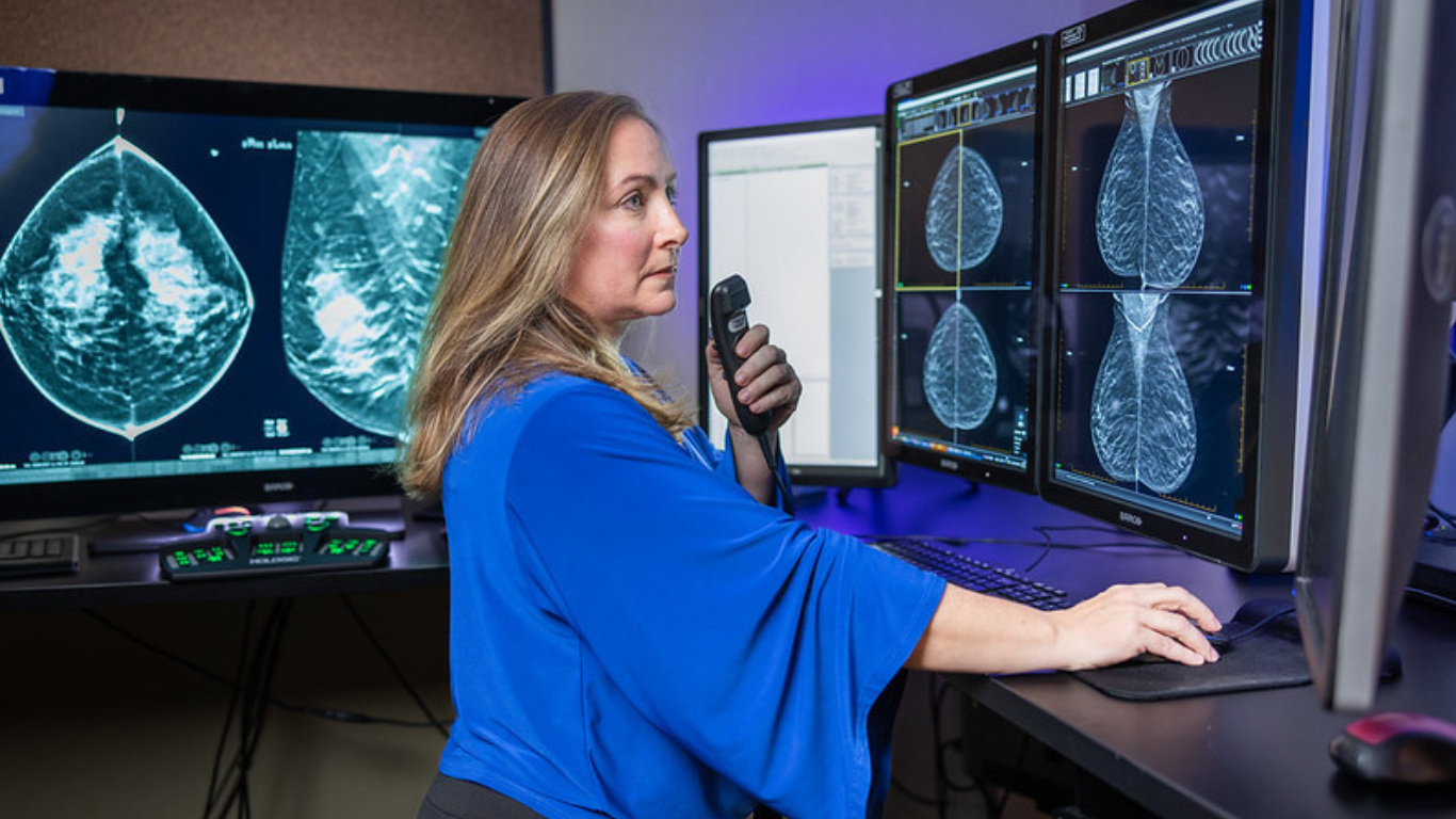
Also called digital breast tomosynthesis, 3-D mammography uses a special approach to enable highly-detailed images of the breast tissue to be captured. RAI is proud to be the first to offer 3D Mammography in Mercer County in an effort to improve breast cancer screening and prevention. 3D mammography is performed in conjunction with traditional mammography; once the 2D images have been made, a special x-ray arm sweeps over the curve of the breast, emitting x-rays as it progresses to make a series of images of thin slices of breast tissue that can be examined individually, viewed as a dynamic interactive animation or combined using special software to produce three-dimensional images of the interior of the breast.
Studies have shown that a 3D mammogram can significantly improve detection of breast cancers, especially those in their earliest stages that might otherwise be missed with traditional mammography alone. The ability to flip through individual images makes it easier to view fine details that could otherwise be hidden behind overlapping tissue. Because images are so accurate and can be viewed in so many different ways, a 3D mammogram also decreases the chances you’ll be called back for additional imaging, an advantage that can decrease the anxiety and worry that often go hand-in-hand with mammography recalls. 3D mammography was approved by the FDA in 2011 and has been widely used since then to help women get more accurate results they can feel confident about.
When is 3D mammography performed?
A screening mammography is used to look for abnormal areas of breast tissue that could indicate cancer in its earliest stages. Unlike diagnostic mammography that looks at a specific area of the breast where abnormal breast tissue has been identified, screening mammography looks at the entire breast to scan for abnormalities. The American Cancer Society recommends women have annual screening mammography exams beginning at age 40 and every year after that, as long as a woman is in good health. For women with certain risk factors for breast cancer, screening mammography may be performed before age 40.
Because 3D Mammography uses advanced technology, there is an added expense associated with 3D mammograms (most insurance companies are covering 3D screening mammograms, we always encourage patients to check with their insurance carrier), and the diagnostic imaging technique also involves a slightly higher amount of radiation compared to the 2D procedure alone. However, because it can detect breast cancer in its earliest and most treatable stages, it’s often recommended for women who have increased risks for breast cancer, including those who have dense breast tissue or a family history of the disease.