What is a breast MRI?
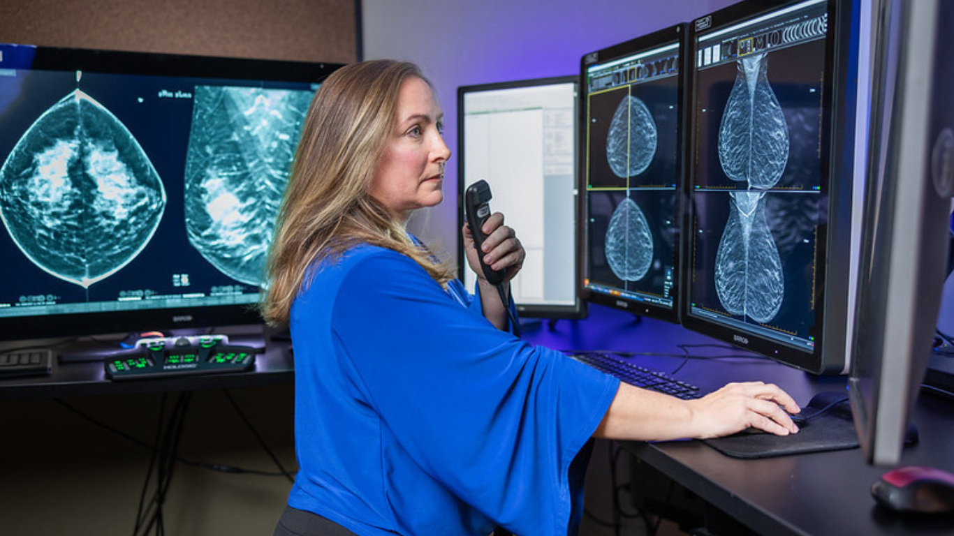
Abreast MRI is a diagnostic imaging procedure that uses magnetic resonance imaging to gather detailed images about the tissues of the breast and the area immediately surrounding the breast. Magnetic resonance imaging uses powerful magnets, radio waves and computer technology to painlessly scan the body’s internal structures, sending a series of images to a computer where they can be viewed and analyzed. These images are like “slices” which, when combined, create a detailed three-dimensional image. Each “slice” contains extremely detailed images of a small portion of tissue that can be viewed individually or with other slices to evaluate a larger area of tissue. While x-rays are primarily used to evaluate harder substances like bones, MRIs can be used to evaluate soft tissues as well. Unlike CT scans and x-rays, MRIs do not use ionizing radiation. Breast MRIs are able to obtain information about the breast tissue that cannot be gathered with mammography or ultrasound; however, it is not a substitute for these diagnostic imaging procedures.
When is breast MRI performed?
Breast MRIs are usually performed as a complement to mammography or ultrasound of the breast to obtain more detailed information about the breast tissue. It may be used to:
- screen women who are at an increased risk for breast cancer, such as those with a strong family history of breast cancer or ovarian cancer, and who could benefit from advanced beast cancer screening
- provide additional, detailed information about abnormalities seen on mammography, and specifically to determine if a biopsy is warranted
- evaluate breast tissue following a diagnosis of breast cancer in order to determine the size of the cancer, how widely the cancer has spread, and whether other cancers are present in the same breast or the other breast
- assess the efficacy of chemotherapy administered prior to surgical removal of the cancer
- provide ongoing evaluation and surveillance of surgical sites in the years following breast cancer surgery
In addition to cancer treatment and assessment, breast MRIs are also performed to evaluate breast implants for signs of rupture or other damage.
How is a breast MRI performed?
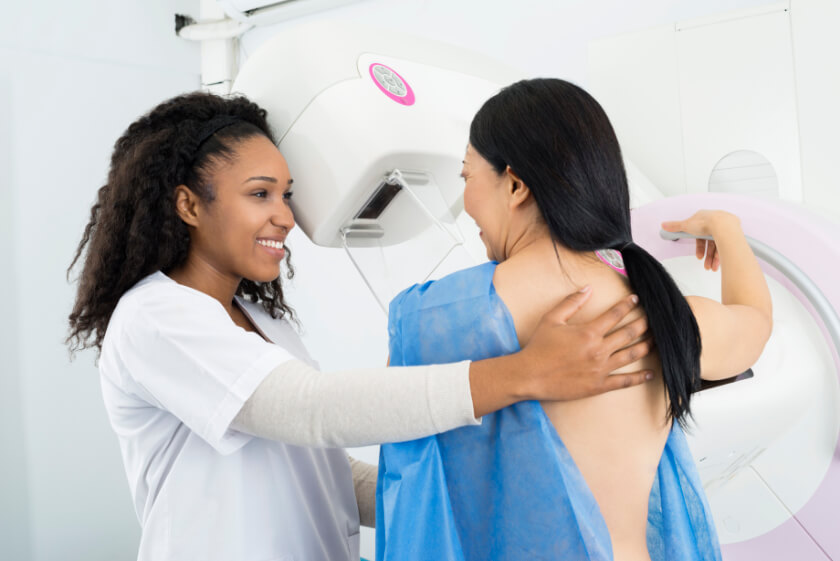
During a breast MRI, you will lie on an exam table on your belly and cushions or bolsters may be used to position you so the clearest images can be obtained. A special exam table is used for breast MRI that allows the tissue to be scanned without being compressed. For most breast MRI procedures, a contrast dye will be used to highlight specific areas of tissue. The dye will be administered through an IV in your arm. Images will be made both before and after the dye is administered. In some cases, an additional test called spectroscopy may also be performed to provide detailed information about the chemicals in your body.
Once you’ve been properly positioned, the table will slowly slide into the MRI device which contains the magnets and other components used to capture the images. During the imaging procedure, the technician will be in another room, but you will be able to communicate using a speaker system built into the device. You’ll need to lie very still to ensure the clearest images are obtained. Most MRI scans take from 30 to 60 minutes to complete. After the MRI is completed, you’ll be able to resume your normal activities.
Eliminating Anxiety with Visual Therapy
We understand that the thought of having to undergo an examination can be stressful and build up unwanted anxiety. At Radiology Affiliates Imaging, our goal is to make every visit comfortable and relaxing for all of our patients so we’ve incorporated visual therapy and nature imagery at both of our imaging facilities. The ceilings of our CT and MRI suites resemble the skies of spring, transforming the rooms into spaces of natural beauty and freshness. The idea of nature imagery, a form of visual therapy, helps you relax during imaging procedures. The minute you enter our suite, you will feel at ease and therefore eliminate anxiety.
Available Locations
Preparation Instructions
-
-
- Please leave your jewelry at home.
- You may have to change into a gown.
- If you are having an abdominal and/or pelvic MRI, you may not eat or drink for 4 hours prior to your appointment. You may take necessary medications with a small amount of water.
- Bring your prescription and insurance card.
- Bring all previous imaging/radiology studies (that were not done at RAI) relating to your current study.
- Please call us at (609) 585-8800 , if you have any of the following:
- Cardiac Pacemaker Artificial heart valve prosthesis
- Eye implants or metal ear implants
- Any metal puncture(s) or fragment(s) in the eye
- Any metal implants activated electronically, magnetically or mechanically
- Aneurysm clips
- Copper 7 IUD
- Penile implant
- Shrapnel or non-removed bullet
- Pregnancy
- Claustrophobia
- Additional questions
-


