Why are biopsies performed?
Biopsies are used to obtain very small samples of tissue for evaluation in a lab, usually using a microscope but sometimes using other techniques as well. Biopsies can be performed for many reasons, including determining if cancer cells are present in the tissue sample as well as testing for other types of diseases and medical conditions. In addition to serving as a very accurate diagnostic tool, biopsies can be used to evaluate the effectiveness of an ongoing treatment so your doctor can decide if the treatment should be continued, stopped or altered in some way for better results. Most biopsies are performed using minimally-invasive techniques using special needles to extract tissue samples without the need for surgery or large incisions.
What is image-guided biopsy?
Image-guided biopsy is a state-of-the-art diagnostic imaging technique used to perform highly accurate biopsies. In an image-guided biopsy procedure, special techniques like ultrasound or fluoroscopy (a kind of “live action” x-ray) are used to ensure the biopsy needle is accurately placed within the target zone – the area from which the sample is to be obtained. By making sure the needle is accurately placed prior to extracting the tissue sample, your doctor can ensure the highest yield of cells for lab analysis, which means your results will be more definitive and your treatment can be highly customized based on your specific needs for superior results and optimal outcomes. Plus, because image-guided biopsy techniques use the most “direct route” to obtain samples, they’re associated with far less risk of bleeding, infection and other potential complications, so you can feel more confident in your care.
What should I expect during the biopsy procedure?
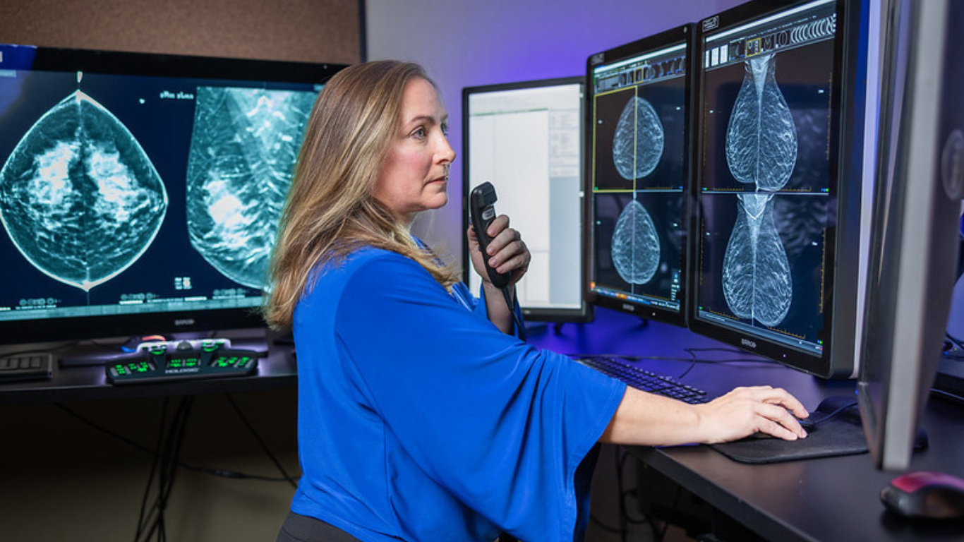
Biopsies can be performed on tissues located throughout your body, but usually, the technique used in an image-guided biopsy is similar, no matter which area is being evaluated. Biopsies are performed using a local anesthetic to numb the site where the needle will enter, and sometimes a sedative to help you relax and minimize discomfort. Diagnostic imaging may be used prior to your procedure to ensure the most accurate entry point for the needle. During the procedure, you may feel some slight pressure or very mild discomfort while the needle is inserted and extracted. As the needle advances toward the target zone, your radiologist will monitor its progress on a computer monitor. Most biopsies take just a few minutes to perform, and once the cells have been extracted, the needle will be removed and a small bandage will be placed over the needle insertion site.
At Radiology Affiliates Imaging, our radiologists specialize in minimally-invasive general biopsy techniques using advanced image-guided techniques for the most accurate results. Our state-of-the-art facility is equipped with the latest medical imaging technology aimed at promoting patient comfort while reducing risks associated with minimally-invasive diagnostic procedures.


