
MRIs and Alzheimer’s: Detecting the Disease Early

About 5.5 million people in the U.S. have Alzheimer’s disease, according to the Alzheimer’s Association, and that number is expected to rise to 16 million by 2050. Alzheimer’s is the most common type of dementia and the 6th leading cause of death, causing more deaths every year than breast cancer and prostate cancer combined. About one out of every 10 men and women 65 years old or older have Alzheimer’s, and while deaths from heart disease have decreased by 14 percent since 2000, the mortality rate from Alzheimer’s disease has risen by 89 percent. In fact, every 66 seconds, someone develops the disease.
Although medical researchers are working feverishly to gain a deeper understanding of the disease that may eventually lead to a cure or even a prevention strategy, today, Alzheimer’s disease is the only top 10 cause of death in the U.S. that can’t be cured or prevented. In the meantime, researchers have uncovered techniques that can actually help identify the disease in its earliest stages and potentially identify people who are at greater risk for the disease. And most of those techniques rely on neurological imaging of the brain using techniques like magnetic resonance imaging (MRI).
Structural Changes in Alzheimer’s Disease
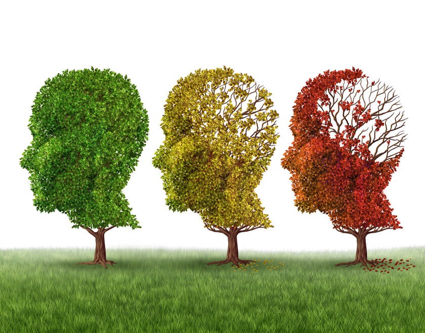
The cognitive and behavioral changes in Alzheimer’s disease are caused by significant changes in the structure of the brain. These changes typically take years to develop, and they begin to occur as long as a decade before any actual symptoms of the disease become apparent. This is considered the “preclinical” stage of the disease. During these months and years, protein deposits build up in the brain, forming abnormal clumps (called amyloid plaques) and tangled fiber bundles called tau. In a healthy brain, tau fibers help maintain the orderly processes in the brain, keeping everything “on track.” But in Alzheimer’s, changes inside the brain cause the fibers to become disorganized and tangled, depriving the brain of important nutrients and oxygen and causing the brain to slowly deteriorate over time.
Neurons stop functioning and neuron-to-neuron connections are lost, contributing to further decline in the brain and in other organs and muscles in the rest of the body. These changes begin in the area of the brain called the hippocampus, a part of the brain that plays a very important role in memory and emotion. As the disease progresses, more areas of the brain are affected, and the loss of neural connections and other structural changes causes the brain to shrink dramatically.
Magnetic resonance imaging provides very detailed images of the brain, helping doctors identify the disease in its very early stages. State-of-the-art 3T MRI imaging is able to provide more detail than traditional MRI, making it very useful in identifying changes in the medial temporal area of the brain where the hippocampus is located. Atrophy in this area is accepted as a diagnostic marker for the early stage of the disease. What’s more, 3T MRI can also aid in ruling out other types of dementia so management strategies can be more effective.
While early diagnosis of the disease does not mean Alzheimer’s can be cured or prevented, it does make it possible for patients, families and other loved ones to implement management strategies early and to plan for eventual care needs. Plus, it may also identify patients who might be good candidates for clinical trials of medications and other therapies currently being evaluated that may help slow the progression of the disease or even provide a cure in the future.
3T MRI imaging is painless, requires no sedatives and does not use radiation. Instead, MRIs rely on very powerful magnets and magnetic fields to align the protons inside your body, measuring fluctuations in those protons as the magnetic field is altered. These fluctuations are sent to a computer and interpreted as images of the brain or other organs. In most cases, the imaging test takes about an hour to complete.
Learn more about 3T MRI and Alzheimer’s disease.
As a leading neuroimaging center in Central Jersey, Radiology Affiliates Imaging is dedicated to providing the most advanced imaging options for patients of all ages. With locations in Hamilton and Lawrenceville, Radiology Affiliates Imaging offers 3T MRI and other diagnostic imaging tests to identify disease processes as early as possible, helping every patient receive optimized treatment based on their needs. To find out more about 3T MRI for Alzheimer’s imaging or to schedule an appointment, call Radiology Affiliates Imaging at 609-585-8800 today.
Related Posts

Brain Imaging Provides a Glimpse into Alzheimer’s Disease
June is Alzheimer’s and Brain Awareness Month and Longest Day, an event held by the Alzhei

Using Imaging, Artificial Intelligence and Genetics to Predict Drug Effectiveness in Personalized Medicine for Patients with Alzheimer’s Disease
Doctors are working towards delivering personalized medicine, an approach in which doctors tailo
Dementia and PTSD Diagnosis: How a 3T MRI Could Help
Mental health issues, such as dementia and post-traumatic stress disorder (PTSD), can significan