Adding Breast MRI Screening to Mammography in Women with Dense Breasts

Mammograms and breast cancer treatments have prevented more than a half million deaths in the United States over the past 30 years, according to the American Cancer Society. Improvements in treatments and screening methods can potentially save even more lives each year.
Breast cancer is a condition in which abnormal breast cells divide rapidly. These cells can invade nearby tissue, spread to lymph nodes, and eventually spread to tissue far from the breast.
Mammography is the gold standard in breast cancer screening because it is non-invasive, relatively inexpensive, and has reasonable sensitivity of 87 percent, which means it does a good job of detecting breast cancer in most women. However, mammograms are not as sensitive for spotting signs of breast cancer in women with dense breasts, so many medical professionals are turning to MRI screening for women with high breast density. They can also use MRI to detect interval cancer, which is breast cancer that appears in between mammograms and often detected with self-examination.
About Breast Density
Breasts consist of fibrous connective tissue, glandular tissue that produces milk, and fatty tissue. Having dense breast tissue, also known as dense breast tissue, means someone has more fibrous and glandular tissue and less fatty tissue than does someone without dense breasts. Dense breast tissue is normal; it is not a medical condition and does not cause symptoms. A woman cannot tell if she has dense breast tissue just by feeling her breasts, and having firmer breasts does not mean she has dense breast tissue.
Dense breast tissue is also common. About half of all women 40 years of age or older have dense breast tissue, according to the Centers for Disease Control and Prevention (CDC), and many women under the age of 40 have high-density breasts.
The density of a woman’s breasts can change over time, but a woman is more likely to have dense breasts if she:
- Is younger
- Is pregnant or breastfeeding
- Is taking hormone replacement therapy
- Has a lower body weight
When radiologists read mammograms, they use a standard system, known as Breast Imaging Reporting and Data System (BI-RADS), to report what they are seeing on the imaging. Published by the American College of Radiology, the BI-RADS system describes the terminology, organization, and classification system for mammography, ultrasound and MRI of the breast. The system allows radiologists reading the mammogram to tell the patient’s physician if there were any unusual findings, and if something was found, if it was not cancer (benign) or cancerous (malignant). In the report, the radiologist may recommend routine screening, follow-up tests, and provide an assessment of breast density.
In a BI-RADS report, the radiologist scores breast density on a scale of A through D, with A being the most fatty and D being the most dense.
A: Mostly fatty – the breasts consist of mostly fatty tissue and contain very little fibrous and glandular tissue; approximately 10 percent of women fall into this category
B: Scattered fibroglandular densities – while the breasts are mostly fatty tissue, a mammogram shows a few areas of fibrous and glandular tissue; around 40 percent of women get this result
C: Heterogeneously dense – many areas of fibrous and glandular tissue are visible on a mammogram; about 40 percent of women have heterogeneously dense breasts
D: Extremely dense – The mammogram shows large amounts of fibrous and glandular tissue; about 10 percent of women have extremely dense breasts
Why is Dense Breast Tissue Important?
Dense breast tissue is important for a number of reasons. Most importantly, women with higher-density breasts have a higher risk for breast cancer.
Dense breast tissue can also make it harder to see signs of cancer in mammograms. In fact, dense tissue can hide cancers in mammograms in about half of all women. This is because glandular and fibrous tissue can look white on a mammogram image – and so can a possible tumor.
Many women with dense breasts must undergo supplemental imaging with either MRI or ultrasound to get an accurate diagnosis; these supplemental imaging tests can help women avoid false positives and avoid needless tissue biopsies.
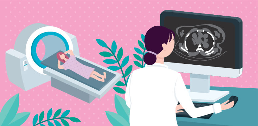
MRI for Breast Cancer Screening
Both breast ultrasound and breast magnetic resonance imaging (MRI) can help a radiologist see through dense tissue, improve a doctor’s ability to identify cancers, and help patients avoid the need for biopsies. Breast MRI offers more specificity than does breast ultrasound, however, which means breast MRI does a better job of helping doctors distinguish which patients have breast cancer and which ones do not.
An MRI is a type of body scan that uses a very large magnet linked to a computer. MRI scans take detailed pictures of tissues inside the breast. It has its drawbacks as compared with mammography, but it also has its advantages. MRI is a more complicated procedure that requires the patient stay in the magnet for about a half hour, so some patients may not be able to tolerate a conventional MRI scan for breast cancer.
However, adding MRI to mammography can help the detection of breast cancer, particularly in women with dense breasts. One study, known as the DENSE Trial, showed that offering MRI to women with very dense breast tissue and normal mammogram results could reduce the patient’s risk of interval cancers by 50 percent.
While mammography is still the gold standard for breast cancer screening, MRI breast cancer screening can help save lives. For more information about breast MRI, contact your doctor or radiology team.
Related Posts

About Irritable Bowel Syndrome
Do you have belly pain, gas, cramping, and diarrhea and/or constipation that comes back again an

Forget BMI – Assessing Muscle Fat May Be Better Indicator of Health
Doctors have warned for years that carrying around extra body fat can increase the risk for seri
Changes in Lung Cancer Screening Rules Saves Lives
Cancer is a leading cause of death. In 2018, doctors diagnosed 18.1 million new cases of cancer