
Could an MRI Help Doctors Determine What Is Causing Your Pain?

Imagine the following scenario: Magnetic resonance imaging (MRI) provides enough detail that the radiologist and orthopedic doctor diagnosed a 50% tear in the peroneal tendon of the right ankle. Without the results from the MRI, the college freshman gymnast would have hurt herself even more. After finishing a dose of steroids to reduce swelling and using an extra-strength pain relieving cream, the anxious gymnast sneaked in a couple of beam routines and base floor tumbling passes at the practice before receiving the results. She knew she was on the travel list but was hoping to earn a spot on the balance beam rotation. Three hours later, however, she was off the line-up for the meet and in a boot to protect the tendon from tearing the rest of the way. She was still traveling for the meet, but she would be watching, not competing.
Sound far-fetched? An MRI can actually provide a more detailed look at an extremity — or even the whole body — than a basic X-ray. That diagnosis lets doctors and their patients know what’s going on in the body to prevent further tears and damage from occurring.
The three-dimensional images produced by an MRI can create in-depth and specific views of bones, tissues, organs, and tendons. Because of the detail from the scans, however, the patient is asked to lie very still for much longer than X-ray. In fact, a basic MRI takes about 30 minutes, but some can last as long as two hours. Because of the loud rattling sound that the imaging machine makes, the patient is given headphones to wear. While some patients are able to sleep during the procedure, many report that the loud clanking sound makes it difficult to rest.
When are MRI Scans Used?
MRI technology has been around since 1977. These machines were so large and expensive they were often transported from hospital to hospital in large trucks. Now, of course, these images are produced throughout the country hundreds of times a day. Radiology imaging can be used in a variety of situations, including:
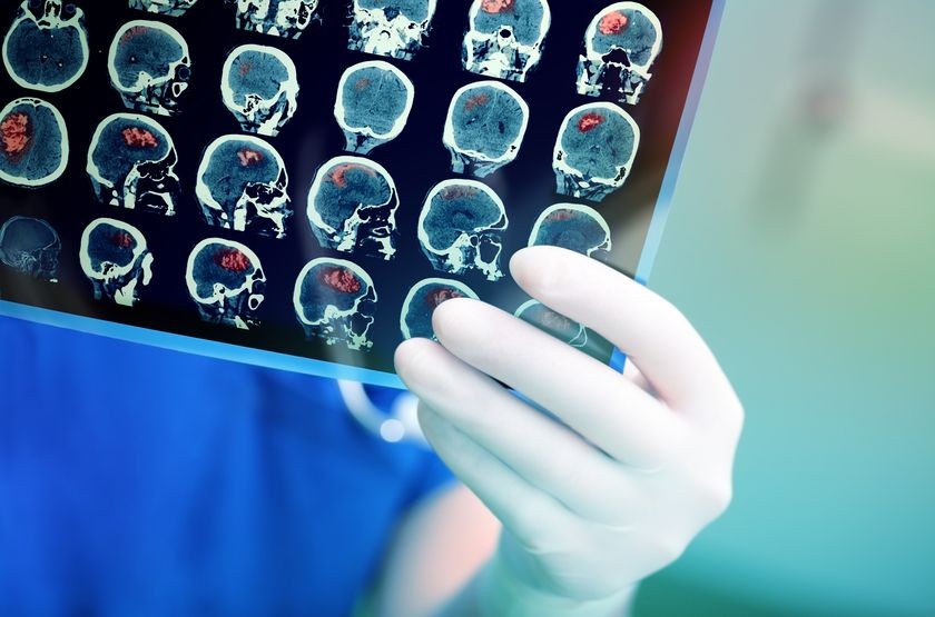
- Head scans. In fact, of the 30 million MRI scans done every year in America, 20% of them are for head scans.
- Spinal conditions and injuries.
- Soft tissue and bone conditions that are not apparent in an X-ray.
- Brain ailments, including tumors and sources of dementia.
- Some kinds of ear, nose, and throat conditions.
- Female pelvic conditions.
- Male prostrate conditions.
What Is the Difference Between an Open MRI and Other MRIs?
An Open MRI refers to the amount of space that is available around the patient’s body. Early scanning procedures using this technology were known for sliding a patient into a narrow opening where the procedure took place. Although a scanning technician is always in the room and able to communicate with the patient, the narrowness of the opening created varying degrees of anxiety in patients. The Open MRI process, however, can vary. While the “openness” of an MRI can refer to any amount of space around the patient, some of the newer machines actually allow a patient to sit or stand during the imaging process.
If a patient is sensitive or concerned about closed spaces (claustrophobic), it might be worthwhile to ask about how “open” the MRI process is. The tunnel scanner often provides the most thorough results, but models that allow the patient to stand or sit are also making advancements in the images they provide.
Is an MRI Scan Right for Everyone?
Pregnant women, obviously, are rarely given MRI scans. Just as they are normally not given X-rays, pregnant women should avoid being exposed to magnetic imaging processes. The first three months of pregnancy are almost always off-limits to magnetic scanning procedures.
Other patients who should not have MRIs are people who have some kind of metal in their bodies. A metal plate in a person’s head, for example, makes that patient unable to have an MRI. Likewise, heart pacemakers and surgically inserted metal joints will keep people from having MRIs.
Related Posts
New MRI Procedure Could Help Doctors Understand Association between Fluid Flow and Brain Tumors
Advances in imaging technology are helping scientists and doctors see how diseases affect the hu
Calcium Scoring, a Crucial Exam at the Lowest Price Around
What is coronary calcium scoring, And How Much Does it Cost? Coronary calcium scoring is a proce

MRIs and Alzheimer’s: Detecting the Disease Early
About 5.5 million people in the U.S. have Alzheimer’s disease, according to the Alzheimer&