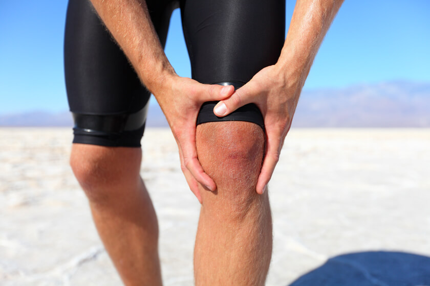
Do You Have an Increased Risk of Knee Injury? A Single MRI Measurement Can Help You Find Out

Knee injuries can sideline professional and amateur athletes alike. The results of a new study suggest that a single magnetic resonance imaging (MRI) measurement can help doctors determine patients’ risk for knee injuries associated with kneecap instability.
The kneecap protects the knee and plays an important role in how the knee bends. It is a small bone situated in front of the knee joint, where the where the thighbone and shinbone, or tibia, meet to form the knee joint.
The kneecap slides up and down in a groove, known as the trochlear groove, when the knee bends. The kneecap can slide out of position to the left or right of the trochlear groove, though, which prevents the leg from bending correctly. The kneecap connects the muscles in the front of the thigh with the tibia, so knee instability can prevent the muscles from moving the leg bones correctly.
Measuring Risk by Measuring the Knee
Researchers have recently discovered that measuring the distance between a bony bump on the tibia, known as the tibial tubercle, and the trochlear groove was a reliable and precise way to predict problems associated with the kneecap moving out of its joint. They also realized that they could measure this distance between the tibial tubercle and the trochlear groove accurately with an MRI.
Ideally, your kneecap sits perfectly in the middle of the trochlear groove, neither too far to the left nor too far to the right. People whose kneecaps sit in the correct spot of the trochlear groove tend to have a relatively short distance between their tibial tubercle and the trochlear groove. Having a greater distance between tibial tubercle and the trochlear groove may suggest that the kneecap is off center, which means it may be more likely to slip out of the joint.
Using MRI to Measure the Tibial Tubercle – Trochlear Groove Distance to Predict Knee Instability
The results of the study, published in Clinical Orthopaedics and Related Research, may help refine how doctors predict knee problems.
“We found that once this measurement reaches 13 millimeters, that individual may be more likely to experience problems with kneecap dislocation,” says John Vairo, lead author and Associate Teaching Professor Clinical Associate Professor of Orthopaedics & Rehabilitation Program Director at Penn State College of Health and Human Development. “That being said, it’s also not the only thing that should be taken into account if a surgeon is considering more aggressive types of treatment like surgery to decrease this distance, which requires significant recovery time.”
The researchers note that kneecap instability issues are common, particularly among women and people who engage in sports. While kneecap instability might not present a problem for those who are not physically active, the condition may require surgery or other aggressive treatment for athletes.
Doctors have been using tibial tubercle – trochlear groove measurements to predict kneecap instability for years, but the medical community has not yet established the best way to take those measurements. Many doctors had been using computed tomography (CT) to measure the distance between the tibial tubercle and the trochlear groove, but research suggests that CT and MRI measure those distances differently. The researchers in the most recent study wanted to test the reliability of the MRI measurements in everyday settings to create a test that a doctor, usually a surgeon considering surgery, could perform one time to determine a patient’s risk for knee instability.
The researchers analyzed the medical records from 131 patients. The 48 patients in the test group had confirmed kneecap dislocations, while the 83 participants in the control group had meniscal tears. Three doctors with different levels of experience used the patients’ MRIs to measure tibial tubercle – trochlear groove distance. None of the doctors knew the patients’ diagnosis when they read the scans.
The researchers found that all three doctors, regardless of experience, were able to accurately measure the distance from the tibial tubercle to the trochlear groove. They also found that the distance tended to be greater in those patients with kneecap instability.
The team of scientists also wanted to take their work a step further by finding a specific distance between the tibial tubercle and the trochlear groove that would accurately indicate the likelihood of kneecap instability. Other studies suggested that doctors could use the measurement as a way to predict instability, but they didn’t give a specific number, in which people with a longer measurement have a risk for kneecap instability and people with a shorter measurement do not.
The researchers in the most recent study came up with a specific number – 13 millimeters. In other words, people whose tibial tubercle is 13mm or further from their trochlear groove have a higher risk of kneecap instability.
In the future, doctors will learn more about using information gained through MRI to predict patients’ risk for kneecap problems and other health issues. For more information about MRI, kneecap instability, or preventive screening, speak with your doctor or radiologist.
Related Posts
Using MRI to Identify Patients with Sarcoidosis at Risk for Bad Cardiac Outcomes
Some people with sarcoidosis experience adverse outcomes that cause severe illness and eve
Cyclical Breast Pain: Common Causes and When to Get a Mammogram
Do you have breast pain that seems to come and go every month? It may be cyclical breast pain. A
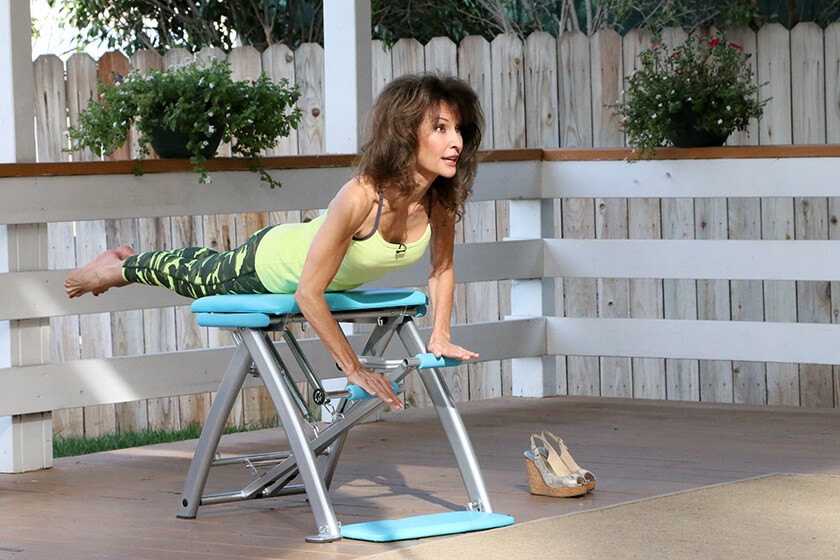
Susan Lucci’s Dramatic Close Call with Heart Disease
Susan Lucci is the picture of health – she’s slim, she exercises and she eats the h