The Role of Radiology in Prostate Care

Prostate care is important to a man’s overall health. Located between the bladder and the penis, this walnut-sized gland secretes fluid that nourishes and protects sperm. The urethra, which is a tube that carries urine from the bladder to the penis, runs through the prostate. Prostate problems can press against the urethra to restrict urination and cause pain. Most men don’t think about their prostates until they have a problem “down there” that causes pain, difficulty urinating, or other symptoms.
Several conditions can affect the prostate. These conditions include benign prostatic hyperplasia (BPH), prostatitis, and prostate cancer. BPH, also known as an enlarged prostate, is a common condition among older men. By the age of 60, about half of all men will have BPH and about 90 percent of men over the age of 90 have an enlarged prostate.
Prostatitis is inflammation of the prostate gland and irritation of the nerves in the area. This painful condition is often the result of a bacterial infection. Prostatitis is common, with about 8.2 percent of men experiencing prostatitis at some time in life. Chronic prostatitis, which lasts longer than 3 months, affects 10 to 15 percent of men in the United States.
Prostate cancer is of special concern. Like other types of cancer, prostate cancer is characterized by the rapid growth and spread of abnormal prostate cells. Other than skin cancer, prostate cancer is the most common cancer in men in the United States. Doctors will diagnose about 191,930 new cases of prostate cancer in 2020, according to estimates by the American Cancer Society. Prostate cancer will claim approximately 33,300 lives this year.
It takes a team of medical professionals a number of medical departments to provide the wide range of services a patient needs when it comes to prostate care. General practitioners are often the primary care providers, as they perform physical exams and order tests. Technicians and pathologists in the laboratory perform the prostate-specific antigen (PSA) blood test that screens for prostate cancer. Surgeons may become involved, as can oncologists, who are doctors that specialize in cancer care.
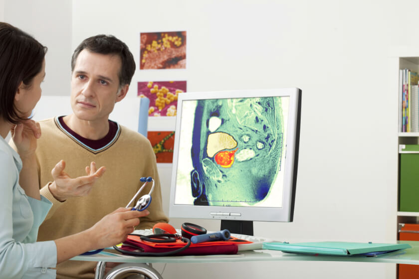
Radiologists and others in radiology are an important part of the prostate care team too. They use ultrasound, magnetic resonance imaging (MRI), and urograms to create images of the prostate gland and surrounding tissue.
Radiology Tests Help Doctors Diagnose Conditions Affecting the Prostate
Prostate ultrasound
Ultrasound is the first line of investigation of prostate problems after the doctor’s examination. Ultrasounds use sound waves and a computer to produce black and white images of a man’s prostate gland. These images can help doctors determine the cause of signs and symptoms, such as an elevated PSA blood test result or difficulty urinating. Ultrasound is non-invasive, safe, and does not use radiation.
An ultrasound can detect disorders within the prostate. Radiologists can use ultrasound to measure the prostate to determine if the prostate gland is enlarged, for example, a finding that would indicate BPH. Ultrasound can detect abnormal growths within the prostate, which may indicate prostate cancer. An ultrasound test can also help diagnose the cause of a man’s infertility.
Radiology also performs ultrasound in ultrasound-guided biopsies of the prostate gland. Biopsies are currently the only definitive way to diagnose prostate cancer, and to differentiate between prostate cancer and BPH. Biopsies are small samples of tissue sent to Doctors use needles to collect a small sample of prostate tissue, and then send the sample to the laboratory for evaluation; they use ultrasound to guide precise placement of the needle.
MRI of the prostate
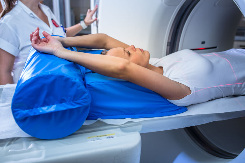
MRI uses a powerful magnetic field, radio waves, and a computer to create images of the structures within the prostate gland. Doctors use MRI of the prostate primarily to determine the extent of prostate cancer, and to determine how far it has spread. Prostate MRI is helpful diagnosing an infection, enlargement of the prostate, or congenital abnormalities. Medical professionals also use MRI of the prostate to evaluate complications after pelvic surgery.
Urogram of the prostate
Formerly known as intravenous pyleogram (IVP), a urogram combines x-rays with special dyes, to create images of the kidneys, bladder, and other organs associated with the urinary tract. Doctors use urograms to diagnose conditions that might be causing symptoms, such as pain or discomfort in the lower back or sides, or when blood is present in a patient’s urine sample. Urograms are helpful in the diagnosis of kidney or bladder stones, cysts, and tumors; these radiology tests are also helpful in diagnosing an enlarged prostate gland.
Armed with the information they gain from radiology tests, doctors can visualize, identify, and diagnose prostate problems, such as enlarged prostate and prostate cancer. Radiology can also help doctors monitor the effectiveness of treatment. For more information about radiology and prostate care, consult with a physician or radiologist.


