Using Imaging Techniques in the Diagnosis and Management of Ankylosing Spondylitis

Ankylosing spondylitis (AS) is a type of arthritis that typically affects the spine, although it can affect other joints. Imaging technology, such as x-rays, ultrasound, and magnetic resonance imaging (MRI) scans can help doctors detect the signs of AS; imaging also helps physicians monitor changes over time and adjust the patient’s treatment plan as needed.
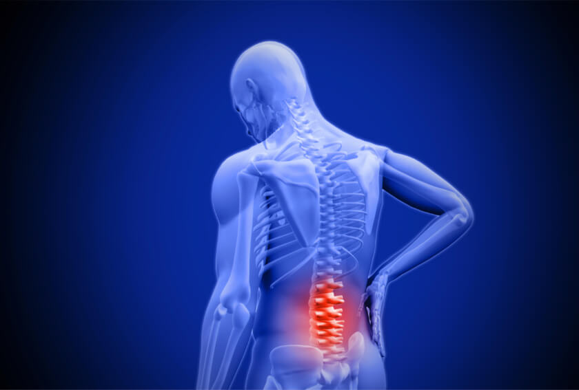
Spondylitis causes inflammation of the joints between the bones of the spine, known as vertebrae, resulting in severe, chronic pain and discomfort. In time, the condition can cause the formation of new bone tissue that can slowly fuse the vertebrae together, in a process known as ankylosis. In other words, the arthritis can eventually cause some of the bones of the spine to fuse together. Fusing the bones together immobilizes the joints of the spine; this can prevent someone from freely moving their back about in all directions.

Using Imaging to Detect and Manage Ankylosing Spondylitis
Doctors can see the effects of AS on the images created by x-rays, ultrasounds, and MRIs. The hallmarks of ankylosing spondylitis are inflammation in the lower back and fusion of the vertebrae of the sacroiliac, which is the part of the lower spine that links to the pelvis.
In addition to using imaging to determine if the patient’s sacroiliac vertebrae are fusing together, a doctor can use x-rays and MRIs to monitor other changes associated with AS. Some patients with AS experience chest pain and stiffness that make it hard to breathe and increase the risk for lung problems, for example, and signs of these lung problems may show up on x-rays and MRI images.
Diagnosing Ankylosing Spondylitis
Diagnosing ankylosing spondylitis involves reviewing the patient’s symptoms, medical history and family history of AS, doing a physical evaluation, and performing blood work and imaging tests. There is no single test for AS, so doctors rely on a body of evidence to diagnose the condition.
Ankylosing spondylitis usually appears before the age of 45, and causes pain in the back and buttocks. Symptoms include chronic back pain that has lasted 3 months or longer; pain often develops over the course of several weeks or months. Back pain and stiffness typically worsens with immobility, with more severe symptoms occurring at night or early in the morning, Symptoms usually go away with physical activity throughout the day. Ankylosing spondylitis can sometimes, but not always, cause chest pain and stiffness.
During the physical exam, doctors look for signs of inflammation by checking for pain and tenderness along the patient’s back, chest, sacroiliac joints, pelvic bones, and heels. Physicians may also check for limitations in the patient’s mobility and for restriction of the patient’s chest cavity. If doctors suspect ankylosing spondylitis, they will also ask if any member of the patient’s family has ankylosing spondylitis or unexplained back pain.
There may be a genetic component to AS. More than half of all people with ankylosing spondylitis have the HLA-B27 gene, as detected with a blood test, according to the Spondylosis Association of America. Specifically, 95 percent of Caucasians with AS test positive for the HLA-B27 gene, as do 80 percent of AS patients from Mediterranean countries and 50 percent of African American patients with AS. Just having the gene does not mean a person will develop AS, though, as 2 percent of people or less born with this gene will eventually develop the condition. Other blood tests can detect inflammation, but cannot definitively attribute the swelling to ankylosing spondylitis.
Imaging Tests Bring Ankylosing Spondylitis into View
X-rays and MRI scans can help confirm the presence of ankylosing spondylitis, and can help doctors and patients make decisions about treatment. Imaging can also help them monitor ankylosis or other changes in the back and alter treatment plans accordingly.
Since AS typically develops in the lower back, doctors often order x-rays of the sacroiliac. X-rays can signs of ankylosing fusion and damage to the joint. An MRI or ultrasound can reveal changes, such as inflammation without joint damage, which do not show up on x-rays. Inflammation can cause a widening or thinning of spaces between the bones of the joint, for example, and increased blood flow to the area. MRIs, ultrasounds, and x-rays can often provide the best evidence to support a diagnosis of ankylosing spondylitis; these imaging techniques can also help doctors monitor changes in inflammation or joint damage.
While there is no cure for ankylosing spondylitis, medications and treatments can ease symptoms and improve mobility. Some medications may even slow the progression of AS. In addition to medications, treatment may include exercise and physical therapy, practicing good posture, and the application of heat and cold to help relax muscles and relieve joint pain.
For more information about ankylosing spondylitis and the imaging techniques used to diagnose and monitor the condition, consult with a doctor or radiology professional.
Related Posts
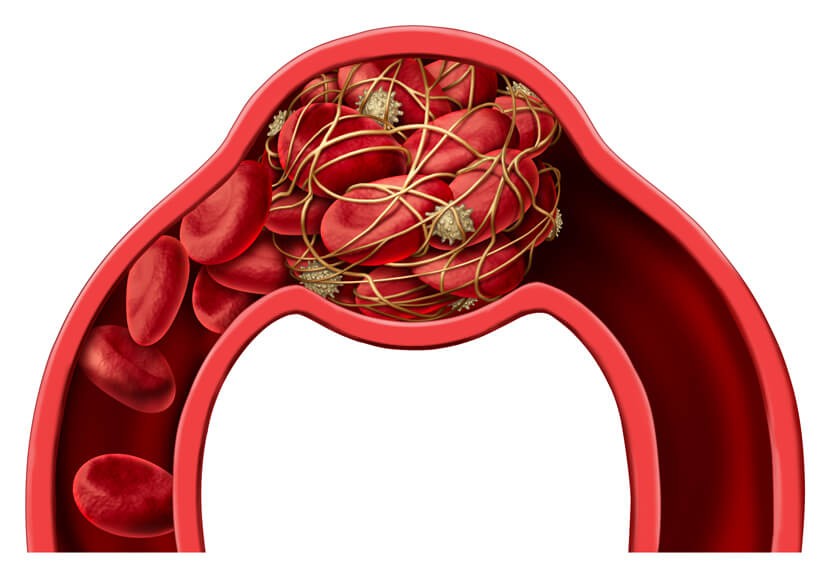
Diagnosing Dangerous Blood Clots with PET/CT Imaging
The results of a new study suggests that PET/CT imaging may do a better job of helping doctors d
FDA Shares New Guidance for Medical Device Manufacturers to Make MRIs Safer
Because it does not use radiation to create images of tissues and organs inside a living body, m

Radiology Preparedness for COVID-19 – Changing the Radiology Department to Improve Overall Care
Coronavirus disease (COVID-19) – and the fight to control it – has continued to change since