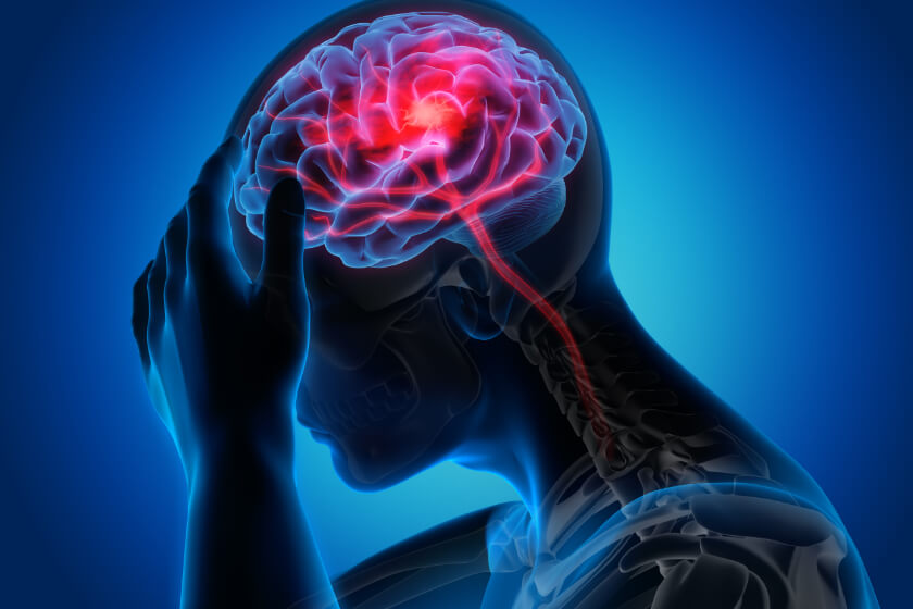
What Happens During a Stroke?

A stroke is a medical emergency that prevents a part of your brain from getting the oxygen- and nutrient-rich blood it needs. More specifically, stroke affects the arteries that carries blood to and within your brain. A stroke can have devastating consequences, including disability and even death. Understanding what happens during a stroke can help you identify the signs and symptoms that you or someone you are with is having a stroke – and the earlier you seek treatment, the better the outcome may be.
Stroke is the fifth leading cause of death in the United States, according to the American Stroke Association. It is also a leading cause of disability in the nation. The Centers for Disease Control and Prevention (CDC) says that, every year, more than 795,000 Americans suffer a stroke. About one in four strokes are in people who have had a previous stroke.
In general, a stroke interrupts the flow of blood to the brain, which prevents brain cells and tissue from getting the oxygen and nutrients they need to function and survive. Even a short interruption of oxygen-rich blood can damage brain cells. Going without oxygen for minutes to hours can lead to the death of brain cells and tissue.
Types of Strokes
There are three main types of strokes, and each happens in a different way.

Ischemic strokes
Ischemic strokes are the most common type, accounting for about 87 percent of all strokes. This type of stroke happens when something narrows or completely blocks the arteries that deliver oxygen-rich blood to the brain.
Blood clots are a common cause of ischemic strokes. Clots can form as the result of atherosclerosis, a condition in which plaque builds up inside the arteries. Consisting of fat, cholesterol, calcium, waste products from cells and clotting agents, the buildup of plaque can clog arteries to prevent blood from flowing well. Plaque can also burst and damage the artery. A clot may form over the damaged area; the clot could break free and travel through an artery in the brain to cause an ischemic stroke.
Hemorrhagic Stroke
Hemorrhagic stroke occurs when an artery leaks blood into brain tissue or completely breaks open in a rupture. The leaked blood causes pressure on the cells in the brain tissue near the artery; excess pressure can damage brain cells. This type of stroke also prevents blood from reaching the other areas of the brain served by the affected artery.
Certain health conditions can cause hemorrhagic stroke. In high blood pressure, for example, blood exerts a tremendous amount of pressure against the inside of arterial walls. This pressure can cause the affected artery to leak blood into the surrounding tissue. Aneurysms, which are balloon-like bulges in an artery, can stretch and burst to cause a hemorrhagic stroke.
There are two types of hemorrhagic strokes, categorized according to the part of the brain the bleeding affects.
Intracerebral hemorrhage affects the tissue inside the brain. This type of stroke occurs when an artery bursts to flood the surrounding brain tissue with blood; it is the more common type of hemorrhagic stroke.
Subarachnoid hemorrhage causes bleeding in the space between the brain and the tissue covering the brain. This type of stroke is less common than intracerebral hemorrhage.
TIA
Also known as a “mini stroke” or “warning stroke,” a transient ischemic attack (TIA) is a warning of a future stroke. It is different from other types of strokes in that the interruption of blood flow to the brain lasts for only a short time – usually five minutes or less. A TIA is the same as other types of stroke in that it is a medical emergency that requires immediate care. Furthermore, a TIA is often a warning sign that a major stroke may occur: more than a third of people with an untreated TIA suffer a major stroke within a year, and as many as 10 to 15 percent of those with untreated TIAs have a major stroke within three months.
TIAs are usually the result of blood clots. Just like an ischemic or hemorrhagic stroke, a TIA is a medical emergency that requires immediate care, as there is no way to tell a TIA from a major stroke.
Effects of a Stroke
The human brain is incredibly complex, with each area of the brain responsible for specific functions throughout the rest of the body. Damage to any area of the brain can cause the loss of normal function in the associated part of the body.
The effects of a stroke can vary widely depending on the type, severity, the number of strokes the patient has had, and where bleeding occurs in the brain.
The brain features three main areas:
- Cerebrum – fills up most of the skull, and is responsible for remembering, problem solving, thinking, feeling, and movement.
- Cerebellum – located at the back of your head, under your cerebrum; controls coordination and balance.
- Brainstem – sits at the base of the brain and controls automatic functions, such as breathing, digestion, blood pressure, and heart rate.
The effects of a stroke may also vary depending on which side of the cerebrum, or hemisphere, they occur. Each hemisphere controls different actions, so a stroke in the left hemisphere may cause different effects than one in the right hemisphere. Furthermore, the right side of the brain also controls many actions on the left side of the body and vice versa, so a stroke on the left hemisphere may cause problems on the right side of the body.
A right hemisphere stroke in the cerebrum can cause left-sided weakness or paralysis, problems with vision, depth perception and memory, difficulty using everyday objects, and behavioral changes.
Effects of a left hemisphere stroke include weakness or paralysis on the right side of the body, and problems with speech, memory understanding language, vision, and reading, along with impaired ability to organize or analyze things, and behavioral changes.
Doctors can detect stroke using imaging that creates pictures of the brain and any bleeding within it. Imaging may include computed tomography (CT) and magnetic resonance imaging (MRI). This imaging helps doctors diagnose stroke, determine its location and severity, predict its effects, and create a personalized treatment plan that helps patients overcome the effects of what happens during a stroke.
Related Posts
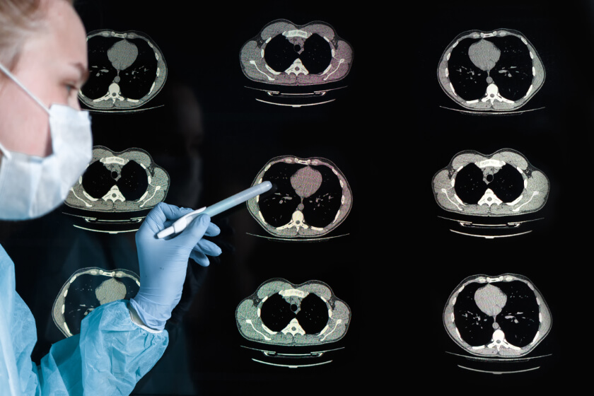
What Does COVID-19 Look Like on a CT Scan?
If you have COVID-19, your doctor might order a CT scan. Have you ever wondered what doctors loo
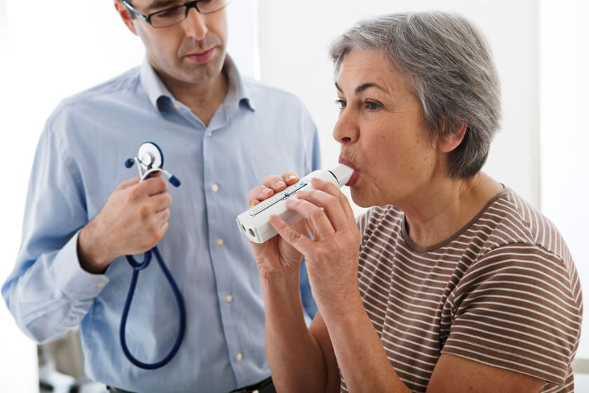
Smaller Lung Airways Increase Women’s Risks for COPD
Many people used to consider chronic obstructive pulmonary disease (COPD) as a condition that on

Study Shows MRI Detects Early Brain Aging for Those with Poor Heart Health
People who have poor cardiovascular health when they are in their 30s have a higher risk f