Why and When to Get Ultrasounds during Pregnancy

Ultrasound provides important information about your pregnancy to you and your health care provider. For example, ultrasound can confirm your pregnancy. This can be helpful when other tests provide inconclusive results. An ultrasound can also reveal whether you are pregnant with twins, triplets, or even more. Ultrasound can help your provider check your baby’s age and growth, which helps your health care professional figure out your due date.
Your health care provider uses an ultrasound to record the movement, heartbeat, and breathing of the fetus. The ultrasound can also help your health care provider determine the amount of amniotic fluid surrounding the baby. The amniotic fluid surrounds and cushions your baby, and helps provide nutrients to your baby, so having the right amount of amniotic fluid is very important.
Your health care provider may use ultrasound to examine your ovaries and uterus. Your ovaries produce and release eggs that, once fertilized, will develop into your baby. Your uterus, or womb, is where your baby grows. Your health care provider can use ultrasound check your baby’s heartbeat, movement, muscle tone, and overall development. The test can also help your health care provider determine if your baby is in the heads-first position right before birth. Being in this position puts your baby in the best possible place for a smooth delivery.
An ultrasound is also helpful for screening to see if your baby has a higher risk of certain health conditions and birth defects. Birth defects are health conditions that change the shape or function of one or more parts of the body, which occur at or before birth. These changes can cause problems in overall health, development, or function.
Since screening only assesses your baby’s risk for health conditions, and does not provide definitive proof that your baby does or does not have the condition, your health care provider may recommend further testing after ultrasound.
Ultrasounds can help health care providers perform other prenatal tests, such as chorionic villus sampling (CVS) or amniocentesis that detect birth defects, genetic diseases, and other problems that can occur during pregnancy. CVS is a procedure in which the health care practitioner removes cells from the placenta using a thin, flexible tube known as a catheter; ultrasound guides the catheter through the cervix to the placenta, which is the tissue that provides nutrients for your baby. Amniocentesis is a procedure in which the health care provider takes a sample of amniotic fluid, which contains cells and fluids produced by your baby, from the sac that surrounds your baby.
Your health care professional may recommend an ultrasound to check for pregnancy complications, such as an ectopic pregnancy, miscarriage, or molar pregnancy. In an ectopic pregnancy, the fertilized egg implants somewhere other where it is supposed to – in the uterus. Miscarriage occurs when an embryo or fetus dies before the 20th week of pregnancy. A molar pregnancy is one in which a non-viable fertilized egg implants into the uterus, but fails to come to term.
When to Get Ultrasounds during Pregnancy
The number of ultrasounds – and the timing of those ultrasounds – may be different for women with certain health issues, such as asthma and obesity. Your health care provider will help you determine how many ultrasounds you should have, and how often you should have them.
By about 6 weeks of pregnancy, it is possible to see your baby’s heartbeat in an ultrasound. It is also possible to predict your baby’s due date, track important growth milestones, determine the number of babies in your womb, and to spot signs of an ectopic pregnancy.
If you are like most healthy women, you will have an anatomical survey ultrasound at 18 to 22 weeks of pregnancy to confirm the normal anatomy and gender of your baby. This test can last 20 to 45 minutes or longer, especially if you are having multiples. This ultrasound is the most thorough checkup your baby will have before delivery. Your health care provider will check your baby’s heart rate during this ultrasound, and look for abnormalities in your baby’s brain, heart, kidneys, and liver. Your health care professional will also count your baby’s fingers and toes.
Your doctor may recommend a transvaginal ultrasound, which is a type of ultrasound that uses a slim, lubricated transducer wand inserted into the vagina. This type of ultrasound provides a clearer image of your baby. In addition to due date, number of babies, and heartbeat, this ultrasound can provide your baby’s “crown-rump length,” which is the measurement from your baby’s head to his or her bottom.
Some women undergo a first-trimester ultrasound, also known as an early ultrasound, before 14 weeks. Your doctor may recommend an early ultrasound if you have certain high-risk pregnancy conditions, such as bleeding, abdominal pain, or a history of miscarriage or birth defects.
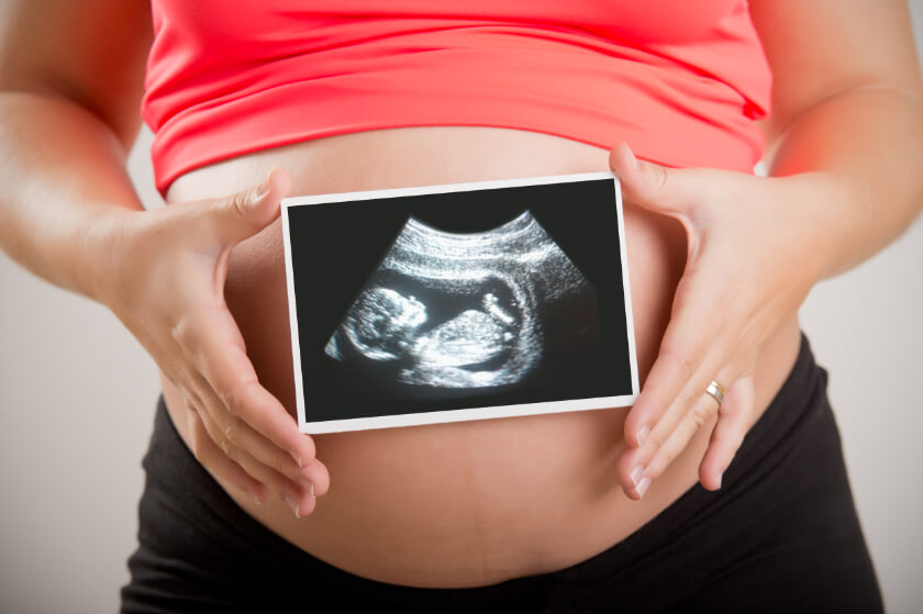
An ultrasound helps provide you with the first glimpse of your baby, even before the big day. For more information about ultrasounds, talk with your health care provider or ultrasound professional.
Related Posts

Low-Dose CT Screening for Lung Cancer Benefits People Who Have Never Smoked
Doctors often recommend low-dose computed tomography (LDCT) screening for lung cancer for patien

Using MRI to Identify Patients with Sarcoidosis at Risk for Bad Cardiac Outcomes
Some people with sarcoidosis experience adverse outcomes that cause severe illness and eve

Is Vaping Bad for My Lungs?
After his father died of smoking-related lung cancer, Chinese pharmacist Hon Lik invented the e-