Chest X-rays Can Help Doctors Predict the Severity of COVID-19 in Young and Middle-Age ER Patients

Like many diseases, COVID-19 can be unpredictable. For example, the coronavirus that causes COVID-19 seemed to affect mainly the elderly in the earliest days of the pandemic, and now it seems to be affecting younger people. Many of these younger patients get sicker now too. Unpredictability can make detecting and treating a disease difficult, though, as doctors need to know what to expect when diagnosing a patient and planning a course of treatment. Because COVID-19 is still relatively new, doctors and healthcare organizations are still working to determine the best tests and treatments for individuals who might have coronavirus.
The Centers for Disease Control and Prevention (CDC) does not currently recommend using chest x-rays to diagnose patients with COVID-19. However, the results of a new study show that these scans can effectively predict which young and middle-aged adults coming to the emergency department with the virus will have a higher risk for developing a more severe illness. The study suggests chest x-ray might also predict which of these patients might require the use of a ventilator to breathe, in a process known as “intubation.”
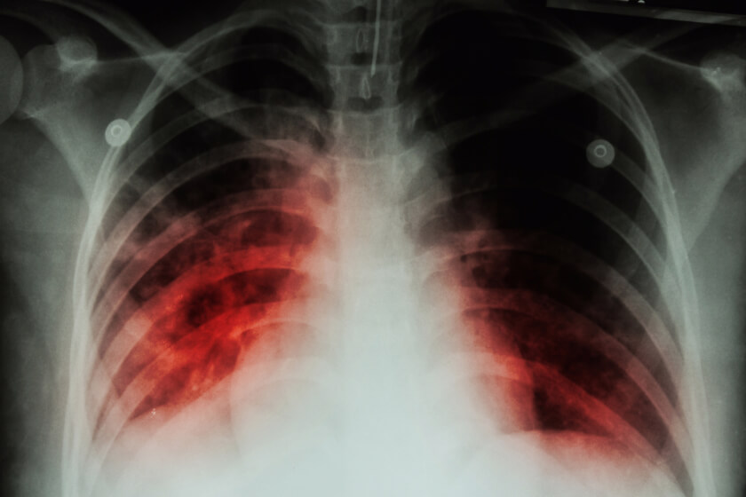
Doctors often turn to radiologists and imaging to help them diagnose a patient and determine the severity of the patient’s illness. Radiologists would interpret images and report areas of concern in the lung where the x-ray was whiter than it should be, a finding known as “opacity.” While the information is helpful for understanding the areas of the lung affected by COVID-19, the researchers in this study wondered if the findings could help doctors predict how serious a patient’s condition would get. The researchers began noticing that most COVID-19 patients had disease in the lower lobes of their lungs, for example, but the patients that had COVID-19 disease in the upper lobes of their lungs seemed to do worse.
The researchers also realized that using a scoring system could help radiologists and referring physicians assess the severity of COVID-19 in patients. Doctors could potentially use the results of this scoring system to make identifying, triaging, and treating these high-risk patients faster. The scoring system would also help doctors get a better understanding of what the chest x-rays results mean for individual patients. To address this, the researchers developed a system that classified the lungs, gave them a score according to the severity of COVID-19 disease, and correlated that score with what the patients actually experienced as their condition improved or worsened.
“This is the first study looking at how we can use chest X-rays from the emergency room to predict how sick COVID-19 patients will get,” said lead author of the study, Danielle Toussie, M.D. “We demonstrate how valuable X-rays can be during this pandemic because, by evaluating disease in different portions of the lungs, we can predict outcomes, which can potentially help appropriately allocate resources and expedite treatment in the most severe cases.”
Researchers Develop System to Use Chest X-rays to Predict COVID-19 Severity in Younger Patients
To assess the usefulness of using chest x-rays in younger patients and to create a scoring system, the team evaluated the records of 338 patients who tested positive for COVID-19 at The Mount Sinai Hospital and Mount Sinai Brooklyn between March 10 and March 26, 2020. Of these, the researchers included the 145 patients admitted to the hospital from the emergency department.
The participants were between the ages of 21 and 50, with an average age of 50. Sixty-two percent of the participants were male. When reviewing the patients’ records, the researchers considered the patients’ race and pre-existing conditions, including asthma, diabetes, high blood pressure, HIV, and obesity.
Each patient in the study underwent a chest x-ray in the emergency department. The researchers examined the patterns of COVID-19 in the lungs of each participant by identifying the opacities and noting the location of the patterns. The scientists divided the COVID-19 chest x-ray patterns into six zones: upper left, upper right, middle right, middle left, lower right, and lower left.
They also created a scoring system to quantify the severity of COVID-19 disease, with zero being the least severe and six being the most severe.
Analysis of the information showed that those with the highest severity scores were 6.2 times more likely to require hospitalization. They also were 4.7 times more likely to be intubated. Thirty-four of the 145 participants scored 0 on the severity score; only two of these patients ended up on a ventilator. In comparison, four out of the five patients with a severity score of 6 were intubated.
Men were more likely to have higher scores, and to have been admitted to the hospital, but males were not more likely to be intubated. Obese participants were more likely to have higher severity scores, and were more likely to require hospitalization.
Study Author Adam Bernheim, M.D., said, “This work is foundational for demonstrating the role of radiology not only in diagnosis, but also in predicting, triaging, and risk-stratifying COVID-19 patients so that those at highest risk for severe disease can be immediately identified from the moment of the very first chest X-ray upon presentation.”
Related Posts

The Role of Radiology in Prostate Care
Prostate care is important to a man’s overall health. Located between the bladder and the

Do You Have an Increased Risk of Knee Injury? A Single MRI Measurement Can Help You Find Out
Knee injuries can sideline professional and amateur athletes alike. The results of a new study s

Cyclical Breast Pain: Common Causes and When to Get a Mammogram
Do you have breast pain that seems to come and go every month? It may be cyclical breast pain. A