Diagnosing Dangerous Blood Clots with PET/CT Imaging

The results of a new study suggests that PET/CT imaging may do a better job of helping doctors diagnose dangerous blood clots than several other methods used today.
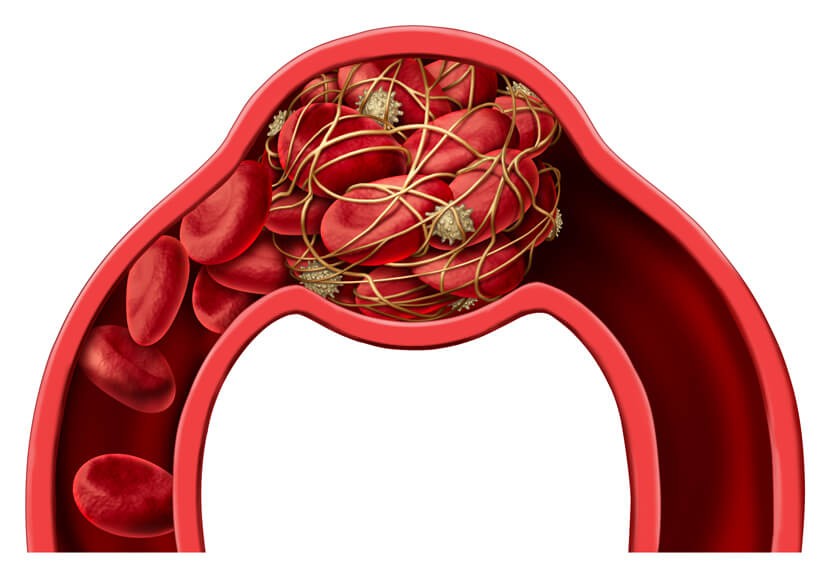
Blood clots are normally a good thing – the formation of blood clots stop excessively bleeding. Sometimes, though, blood clots can pose a health hazard and can even be life threatening. Doctors refer to blood clots as emboli or thromboemboli.
These blood clots can stick to the inside of a blood vessel to stop blood from flowing through it. Clots can also break free from the vein wall and travel to different parts of the body to restrict blood flow there. Doctors refer to these types of clots in general as venous thromboembolism (VTE).
Specifically, VTEs are blood clots that start in a vein. There are two main types of VTE:
- Deep vein thrombosis (DVT) – a clot in a deep vein, typically in a leg, although they can develop in the arms or in a vein elsewhere in the body.
- Pulmonary emboli (PE) – a clot that breaks free and travels to a lung to partially or completely block the blood flow to that lung.

VTE affects between 300,000 and 600,000 people in the United States each year, according to the American Heart Association.
VTE often causes non-specific symptoms that can vary significantly between people, which can lead to delayed or inaccurate diagnosis of the condition. Timely and accurate diagnosis is critical for prompt treatment and a good outcome.
Detecting and Diagnosing Dangerous Blood Clots
To detect and diagnose VTE, doctors currently use ultrasound and two types of computed tomography (CT): CT venography and CT pulmonary angiography. CT venography is a type of CT that allows doctors to create images of veins, particularly in the legs. CT pulmonary angiography uses CT to create images of the pulmonary arteries that carry blood from the heart to the lungs.
Conventional testing with ultrasound and CT safely and effectively detects blood clots, but they are somewhat limited in their ability to identify blood clots in veins below the knees. Furthermore, these tests may not be able to distinguish between older, more stable blood clots from newer, potentially more unstable emboli.
Incorrect diagnosis of VTE leads to unnecessary treatment with anticoagulants to thin the blood and dissolve the clot, which increases risks and costs to the patient. False-negatives puts patients at high risk of potentially fatal PEs.
While ultrasound, CT venography and CT pulmonary angiography are excellent diagnostic tools, they may not be able to distinguish old blood clots from new, potentially unstable emboli. This research suggests a different approach to imaging blood clots by using positron emission tomography (PET) scans and a new type of tracer.
PET scans are a type of nuclear medicine, which means they use small amounts radioactive tracers to help create images. Various cells, such as blood cells, absorb or “uptake” the radioactive materials; the PET scan then detects the tracers to create an image.
PET scans use a number of different radioactive tracers, depending on what the doctor is trying to image. In this study, the researchers found that 18F-GP1 tracer could detect the difference between blood clots and the rest of the blood. Furthermore, the tracer showed the uptake of the tracer in the veins of the lower legs not detected with other types of imaging.
New Tracer Shows Promise for Safe, Effective Diagnosis of VTE
The results of this new study shows that using a specific tracer with a PET scan or CT test could help doctors detect, characterize and track any newly formed clots that have the potential to become a DVT or PE. A research team from Asan Medical Center of the University of Ulsan College of Medicine in Seoul, Republic of Korea, participated in the study. They published their findings in The Journal of Nuclear Medicine.
The research team enrolled 10 patients with DVT and 10 patients with PE into the study. First, the researchers performed ultrasound, CT venography and CT pulmonary angiography. The results of the tests identified PE in five DVT patients and DVTs, and identified DVTs in seven PE patients. Altogether, the tests indentified DVT in 76 veins of 17 patients, and found PE in 245 pulmonary arteries in 15 patients.
Next, the participants underwent PET/CT scan using radioactive tracers, also known as radiotracers, which makes the veins and blood clots easier to visualize. The authors of the study reported that all of the participants tolerated the test well, and that none of the patients experienced adverse effects.
Using the PET/CT and the 18F-GP1 tracer, the research team identified blood clots in 10 patients with acute DVT and 10 with acute PE. The tracer revealed DVT in 16 of the 17 patients and PE in all 15. The testing reported one false-negative, which means testing failed to find a blood clot in the calf muscle of one patient who was suffering shortness of breath and chest discomfort a week after undergoing knee replacement surgery.
While research is still in its early stage, studies like this show that PET/CT scans using 18F-GP1 tracers can accurately locate emboli and provide information on the risk the clot presents. The information provided in this study can eventually change the way doctors treat patients they suspect of having dangerous blood clots.
Related Posts

3D Mammography Provides Special Benefits for Older Women
Mammography is an effective breast cancer screening method for women ages 65 and older, but the

Doctors Use CT Scans to Diagnose and Manage Lung Disease Associated with Vaping
Electronic cigarettes, also known as e-cigarettes or “vaping,” have been making the

Do You Have an Increased Risk of Knee Injury? A Single MRI Measurement Can Help You Find Out
Knee injuries can sideline professional and amateur athletes alike. The results of a new study s