History of Radiology

Radiology is an important part of medical care today. The field developed relatively quickly in the 20th Century, and thanks to computer technology and artificial intelligence (AI), is still advancing.
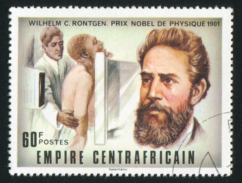
Radiology began in 1895, when Wilhelm Rontgen accidentally discovered x-rays, which is a type of radiation that can penetrate most solid objects. The German physicist was studying what happened when he passed an electrical current through different gases at low pressure. To perform this experiment, he hooked up electrodes to a glass cathode tube and applied voltage to the gas; doing this causes a beam of electrons, known as a cathode ray or electron beam, to go from one end of the tube to the other. Cathode rays glow a green color when they strike the walls of a glass tube.
Rontgen’s ultimate goal was to determine if the cathode rays could pass through glass, so he covered the tube in heavy black paper. The scientist was surprised when the rays not only passed through the glass, but they also passed through the paper, projecting the incandescent green light onto a fluorescent screen sitting a few feet away. Through experimentation, Rontgen learned that the mysterious light could pass through almost all solid objects. Because he did not know what the rays were, he called them x-rays, where the x means “unknown.”
He also found that he could create images of objects put between the cathode rays and a photographic plate. The rays would create images of objects of different thicknesses. At one point, his wife put her hand over the photographic plate; the x-ray image showed the bones of her hand and the ring she was wearing, surrounded by a faint outline created by her flesh. It was the first x-ray of a human body part.
Within a year of the first x-ray, a Glasgow hospital opened its first radiology department, which produced the first pictures of a penny lodged in a child’s throat and of a kidney stone. Soon thereafter, an American doctor used x-rays to trace food as it made its way through the digestive tract. Sometime around 1900, Thomas Edison developed fluoroscopy that creates something similar to an “x-ray movie” by passing an x-ray beam through a body part as the person moves; the fluoroscopy machine then transmits the beam to a TV-like monitor so that doctors can see body parts and movements in detail. In 1918, George Eastman introduced film as an alternative to the glass photographic plates scientists previously used to capture x-ray images.
Introducing Ultrasound, PET, CTs, and MRIs
In the decades that followed, scientists began using other means to create images of tissues inside the human body. Scottish obstetrician Ian Donald developed the first ultrasound for medical use in 1958, for example, and began using it to observe the health and growth of fetuses, and to study lumps, cysts, and fibroids. Donald also worked with engineer Tom Brown to develop the first portable ultrasound for patient use. Ultrasounds use a small handheld device, known as a transponder, which sends sound waves through human tissue. The sound waves bounce off denser tissue back to the transponder to create an image.
In 1961, James Robertson built the first single-plane positron emission tomography (PET) scan. This type of imaging uses a special dye containing radioactive tracers swallowed, inhaled, or injected into the patient’s vein. The PET scan machine detects the radioactivity to create images. Doctors often use PET scans to detect early signs of cancer, heart disease and brain disorders.
Godfrey Hounsfield built the first prototype of a computerized tomography (CT) machine in 1971. A CT combines x-rays with computer software to make three-dimensional (3D) cross-sectional images of the human body. Doctors performed the first successful medical scan using a CT on a live patient that same year.
In 1973, Paul Lauterbur developed a way to generate two-dimensional and three-dimensional magnetic resonance images (MRIs). MRIs use strong magnets to align proton atoms in water molecules inside the body; when the radiology technician turns the magnet off, the protons resume their normal spin and emit energy. Different tissues in the body emit different energy. MRI scans detect this energy to create pictures of the various body tissues.
Doctors around the world continue to use these imaging techniques today. Throughout the following years, researchers have learned how to make x-rays, ultrasounds, CT scans and MRI scans safer and more effective. Today, doctors rely on these radiology techniques to diagnose diseases and guide patient care.
For more information on the history of radiology, or to learn more about how radiologists create images of the human body, consult with a doctor or radiology professional.
Related Posts

Understanding the Different Types of Breast Cancer
There are many types of breast cancer, and many ways to describe them. Reviewing the various ter

American Society of Breast Surgeons (ASBrS) Issues New Mammography Recommendations: Reversing the Trend Towards Later Mammograms
Women at average risk for breast cancer should begin annual mammograms at the age of 40, accordi

November is COPD Awareness Month
Every November, healthcare professionals and patients come together to share information about c