These 3 Imaging Tests Can Help Determine Your Risk of Stroke

Every year in the U.S., nearly 800,000 people suffer a stroke, according to data from the CDC, and more than 600,000 of those are first-time strokes. What’s more the CDC says about 130,000 people die each year as a result of strokes, which are also the leading cause of long-term disability in the U.S.
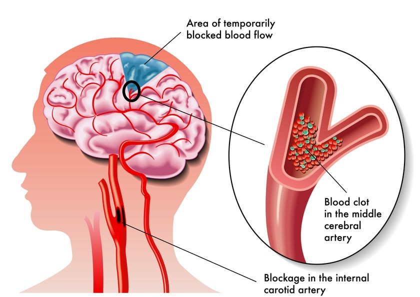
Most strokes – about 85% – are ischemic strokes, occurring when a blocked or narrowed blood vessel interferes with normal blood flow to or in the brain. The remaining 15% are hemorrhagic strokes, occurring when blood vessels burst or leak blood inside the brain. Once blood flow is blocked or significantly slowed, brain tissue is deprived of oxygen and other nutrients, and tissue death can occur within moments. In fact, the National Stroke Association says that for every minute a stroke goes untreated, as many as 1.9 million neurons can die, and that can mean long-term or even permanent loss of mobility, speech and cognitive function.
Strokes can occur in anyone, but they’re more common among people with a personal or family history of stroke, heart disease, hypertension or high cholesterol, and they also become a lot more common as we get older. May has been designated National Stroke Awareness Month, which makes it the ideal time to learn about the steps you can take to limit your risks and hopefully prevent a stroke from occurring.
Stroke Prevention: Steps You Can Take
Lifestyle changes like losing excess weight, exercising more, limiting sodium intake and eating a healthy, low-fat diet are all integral parts of any stroke prevention program, and so is knowing your risk factors. Some risk factors can be gleaned from a personal and family health history, including factors like heart disease, high blood pressure and high cholesterol. But others can only be identified with diagnostic imaging like ultrasound and MRI. And that’s where routine stroke screening can play an important role. To date, there are three primary methods used to help identify these risk factors, and they’re all completely noninvasive and painless:
Carotid Ultrasound
Ischemic strokes are most commonly caused by a buildup of sticky plaques in the carotid arteries, the arteries that carry the largest supply of blood to the brain. There are two carotid arteries, one on either side of the neck. In stroke prevention, carotid artery screening is performed with an ultrasound evaluation to “see inside” the arteries, looking for areas of narrowing and other indicators associated with an increased risk of stroke. During a carotid ultrasound, the doctor or technician will assess the blood flow in the artery, measure the speed of blood flow and determine the diameter of the artery. Ultrasounds can also be used to measure the thickness of the inner layers of the arteries (called the intima media) and to provide detailed information about plaque buildup.
Carotid Artery MRI
More recently, researchers have been evaluating the use of MRI in identifying arterial plaques that are more “vulnerable” – that is, more likely to cause strokes. In one recent study, researchers concluded carotid MRI can “accurately predict” strokes even in those with no history of heart disease.
3T MRI
In addition to assessing the carotid arteries, MRI – specifically 3T (or 3 Tesla) MRI – can be used to evaluate the health of blood vessels inside the brain. 3T MRI technology takes advantage of a higher field strength to obtain very detailed images in less time than standard 1.5T MRI scans. The increased signal used in 3T MRIs helps doctors identify underlying stroke mechanisms like plaque buildup, as well as determining the extent and cause of those plaques, and it can even be used to detect complex plaque formations and identify the components of those plaques.
Signs of Stroke
Stroke screening plays an important role in identifying risk factors and physiologic changes that can significantly increase the risk of having a stroke, but no stroke prevention blog post would be complete without a review of the most common signs of stroke. Stroke symptoms occur very suddenly and can include:
- Weakness or loss of sensation in the arm, leg or face, especially when symptoms affect one side only
- Confusion or difficulty understanding speech
- Difficulty speaking or swallowing
- Vision problems
- Dizziness, balance problems or problems with coordinated activities like walking
- Severe headache with no known cause
The American Heart Association suggests using the acronym “F.A.S.T.” as a quick way to recognize the most common signs of stroke so you can get help right away:
- F – Face drooping or numbness, especially on one side; does the person’s smile look lopsided?
- A – Arm weakness; is one arm weak or numb, or does it drift downward when lifted?
- S – Speech difficulty; is the person’s speech hard to understand? Does it sound slurred? Can the person repeat a simple sentence?
- T – Time to call 9-1-1; if any of these symptoms occur, call 9-1-1 immediately and take note of the time when the symptoms first appeared.
Are you at risk for stroke?
Stroke screening is recommended for anyone with two or more known risk factors for stroke, including hypertension, high cholesterol, tobacco smoking, family history of ischemic stroke, or a first-degree relative who was diagnosed with atherosclerosis (“hardening” of the arteries) before age 60. Screening may also be recommended for people with chronic diseases like cardiovascular disease and diabetes. To find out more about screening techniques and whether stroke screening should be part of your healthy living strategy, contact Radiology Affiliates Imaging, the leader in state-of-the-art carotid ultrasound, carotid MRI and 3T MRI services for patients throughout Central New Jersey. Call RAI 609-585-8800 and schedule a consultation today.
Related Posts
Scientists Use MRI Scanners to Detect ‘Brain Rust’ in Schizophrenic Patients
Using a new kind of MRI measurement, neuroscientists recently discovered that excessive oxidatio
My Battle with Epilepsy and How a 3T MRI Made a Difference
When I was diagnosed with epilepsy, it surely came as a shock. I had a basically normal lif

Using MRIs to Detect Autism in Babies
Is it possible to know as early as infancy when a child is on the autism spectrum? This is a top