What to Expect During an Extremity MRI

An extremity MRI is a type of scan used specifically for diagnostic imaging of the arm, leg, hand, or foot. The machine uses radio waves and a magnetic field to generate images of the inside of the extremity in order to diagnose problems with the muscles, bones, joints, nerves, or blood vessels.
What conditions can an extremity MRI diagnose or assess?
Some conditions include:
- Degenerative joint diseases
- Nerve damage
- Bone infections
- Tumors
- Fractures
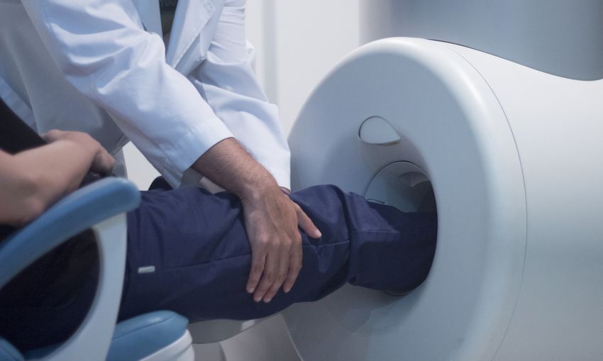
Unlike a traditional MRI machine in which you need to lie still on a table for up to 90 minutes while the scanner takes a series of pictures, extremity MRI scans are much more comfortable. For this kind of MRI exam, you will simply sit in a comfortable chair and place your arm or leg in a small opening in the machine. Your head and torso will remain outside of the scanner, eliminating the claustrophobic feeling many patients experience during traditional MRI exams.
If your hand or arm is being scanned, you will most likely be permitted to stay fully clothed, though it is recommended that you wear a loose top and remove all jewelry. If your leg or foot is being examined, you may be required to wear a hospital gown.
Your doctor will ask you to sit perfectly still during the procedure, though he or she may have you hold your limb in a specific position for a set amount of time. You will hear thumping, tapping, or snapping noises as the magnetic fields shift in the machine.
An extremity MRI will usually take about half an hour, though some may last longer. After the procedure is complete, you will be able to return to regular activities.
What are the benefits of an extremity MRI?
Many people find standard MRI machines to be uncomfortable. Some patients feel anxious and claustrophobic inside the tube. The extremity MRI eliminates that problem, allowing patients to sit comfortably during the exam while a specific body part or “extremity” is being scanned.
Extremity MRIs use the same technology as standard MRI machines to create detailed images of the body for diagnostic purposes. MRI scans are noninvasive, totally painless, and, unlike x-rays, they do not emit any radiation.
Related Posts

MRI and Claustrophobia: Finding Ways to Cope
Claustrophobia is an irrational fear of being enclosed and having no way to escape. Unfortunatel