Your Doctor Ordered a Screening Mammogram: Now What

A yearly screening mammogram brings anxiety for many women, in part, because they don’t have all the information they need to understand how the test works and why it is so important. It’s that time of year again. Your doctor has ordered a screening mammogram for you, so now what?
Why Get a Screening Mammogram?
Your annual screening mammogram is a diagnostic tool able to find the lumps that cannot be felt or are missed during a breast exam. A mammogram is a type of x-ray done that provides doctors with an image of your breast tissue. It is a test that will also detect the presence of microcalcifications, which are tiny deposits of calcium that may indicate cancer. These are all important reasons to get a screening mammogram.
Although the American Cancer Society recommends annual screening mammograms for women 45 years and older, our radiologists follow the American College of Radiology’s recommendation of 40 years and older. The doctor may recommend a mammogram at a younger age if you fall into a risk category like there is a family history of breast cancer or you have never been pregnant.
Why is It Called a Screening Mammogram?
It’s called a screening mammogram because it is ordered to detect any signs of a problem. This is different than a diagnostic mammogram, which a doctor might recommend to investigate a suspicious lump or a complaint like breast pain. A diagnostic mammogram might also be used to get a closer look at something found during the screening test.
Today enhanced technology produces much better images for mammograms, so doctors get a clear picture of the breast tissue. For example, a digital, or full-field, mammography creates a digital image, which is more defined than the conventional film pictures. Computer-aided detection, or CAD, systems do searches of the digital images and bring them to the attention of the radiologist. 3D mammography, also known as tomosynthesis, is another diagnostic imaging tool that allows radiologists to take detailed images with more clarity than its digital and film mammography predecessors.
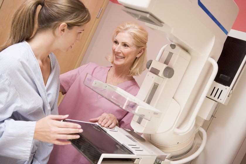
What Happens During a Screening Mammogram?
If this is your first screening mammogram, you might wonder what to expect? When you enter the facility, a technician will have you change into a gown for better access to your breasts during the imaging. The test starts with the technician placing one breast on a platform and making any necessary adjustments to get the best picture. This may mean raising or lowering the platform to fit your height and asking you to shift your body around.
As the test proceeds, your breast is pressed against the platform, so you might have some discomfort, but it doesn’t last but a few seconds. The compression allows the x-ray machine to penetrate the dense breast tissue to get a clear image. The combination of the platform and plate pressing against your breast keeps you from moving around, too, which might blur the picture.
When the technician is ready to take the x-ray, you will hold your breath to prevent chest expansion. The image takes just a few second to complete. When you are told to breathe again, the technician will look at the image to make sure it is clear and readable before shifting your position. This allows for a second image if necessary. When one side is complete, the process is repeated for the other breast. The screening test is over in less than 30 minutes.
Who Screens the Mammogram?
After the test is complete, a radiologist examines the screening images looking for:
- Calcium deposits in the ducts or tissue – This can be an indicator of cancer
- Masses or lumps that need biopsy or further investigation to rule out cancer
- Areas of the image that appear to be asymmetric
- An unidentified dense area on the breast
If you have had a previous screening mammogram, they will compare the two images looking for changes in the breast tissue that require further evaluation.
What If They Find Something?
For many women, the fear doesn’t come from the test itself, but what it might show. Only about 8 percent of screening mammograms detect anything that needs further testing. In addition, a lump or suspicious area in the breast tissue does not automatically mean cancer.
In most cases, the doctor will request a follow-up diagnostic mammogram to determine if there is really a problem. The physician may also recommend an ultrasound of the affected breast to get a different view of the suspicious area.
Screening mammograms can be distressing for some women, but they are also the most comprehensive way to rule out a disease that kills more than 40,000 women a year.
Related Posts

Skipping Your Mammography Exam Could Increase Your Risk of Death from Breast Cancer
Women skip mammograms for a wide variety of reasons. Many women forgo their routine mammogram be

New Study Shows the Value of Getting a Second Opinion When You Receive a Diagnosis of Breast Cancer
When they receive a diagnosis of breast cancer, many women and their doctors want to begin treat

Study Shows Abbreviated Breast MRI may be Best Additional Screening Option for Women with Dense Breasts
Traditional mammograms are less effective at detecting breast cancer among women with dense brea