Dementia and PTSD Diagnosis: How a 3T MRI Could Help

Mental health issues, such as dementia and post-traumatic stress disorder (PTSD), can significantly decrease the quality of life in the patients who suffer them. In many cases, the signs and symptoms of these mental health issues are quite similar; this can lead to incomplete or even inaccurate diagnoses. There is an association between dementia and PTSD – with the presence of one condition increasing the risk of the other – and this close association can make it difficult for doctors to diagnose patients with these common mental health issues. Fortunately, advanced imaging can help.
The Association between Dementia and PTSD
One study shows that veterans with PTSD were at much higher risk of developing dementia than were veterans without the disorder. The study also shows that the risk varies according to the medications the study participants were taking. Those taking psychotropic medications, such as antidepressants, anti-anxiety drugs and mood stabilizers, were at higher risk for developing dementia than were those who did not take these medications.
Older adults and veterans with dementia may exhibit more symptoms of PTSD according to the Veterans Administration (VA).
Conversely, PTSD may be a risk factor for dementia. In fact, results from a large VA cohort study show that people diagnosed with PTSD were nearly twice as likely to develop dementia that were those not diagnosed with the disorder.
About Dementia and PTSD
Dementia is relatively common in older adults, affecting about one in six women and one in ten men, according to the Institute for Dementia Research & Prevention at Pennington Biomedical Research Center at Louisiana State University System.
An estimated 7.8 percent of people in the United States will experience PTSD at some point in life, according to the Nebraska Department of Veterans’ Affairs. Approximately 30 percent of people who have spent time in a war zone will develop PTSD.
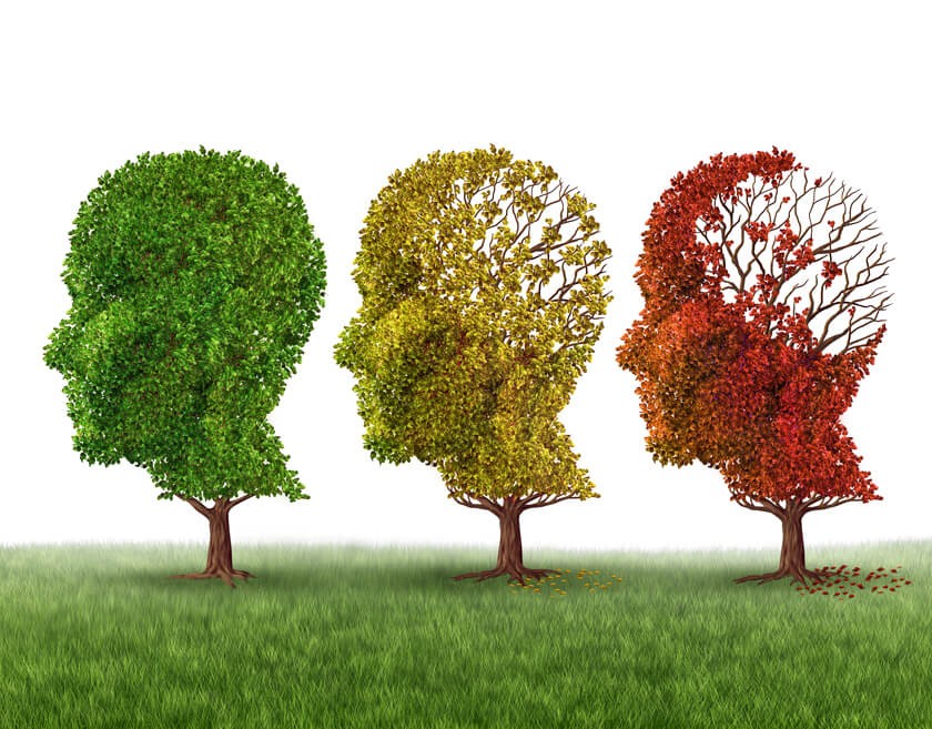
Dementia is not a specific disease, according to the Alzheimer’s Association, but rather an umbrella term that describes a wide range of symptoms.
The human brain has many distinct regions, and each is responsible for carrying out different functions, such as memory, judgment or movement. When brain cells within a certain region sustain damage, the brain cells in that region have trouble performing their appointed tasks. Damage to cells in the region responsible for memory causes memory problems, for example. In other words, the symptoms correspond to the area of the brain affected by cell damage.
Dementia is the result of damage to the brain cells. The damage prevents the brain cells from communicating with each other in ways that affect how a person thinks, feels, and behaves.
Dementia is a degenerative disease, which means the changes to the brain and the symptoms they cause get worse over time. One of the most difficult factors in diagnosing dementia is that it is also heterogenic, which means the disease can progress differently in each person with the condition. Dementia may develop quickly in one person and slowly in another, for example, and the disease can cause different changes in one person’s brain than it does in another person’s gray matter.
Doctors are not yet sure what causes Alzheimer’s and other forms of dementia, but they suspect that plaque and tangles have something to do with cell death and tissue loss in the brains of people with Alzheimer’s disease. Plaques are abnormal clusters of protein fragments that build up between nerve cells in the brain. The dead and dying nerve cells in the brain of an Alzheimer’s patient contain tangles, which contain twisted strands of another protein, known as tau.
When doctors use a microscope to view and compare brain tissue from patients with Alzheimer’s disease and tissue from patients with normal brains, they can see that brain tissue from Alzheimer’s patients contains substantially fewer brain cells and synapses. They can also see the presence of tangles.
Not all types of dementia cause tangles and plaque, though. Research shows that MRI can help doctors differentiate between Alzheimer’s disease and other types of dementia.
MRI for Dementia
MRI is often considered the preferred imaging examination for Alzheimer disease because the test allows for accurate measurement of the 3-dimensional (3D) volume of those brain structures associated with the disease, particularly the size of the hippocampus and other related regions.
Neuroimaging with MRI is also useful for excluding other forms of dementia that may be reversible, such as brain tumors, and for excluding other causes of dementia, such as cerebrovascular disease.
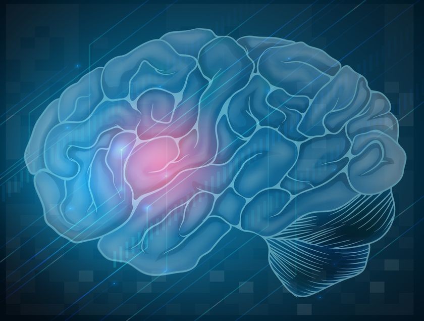
Traumatic stress, like the stress that brings about PTSD, can have a broad range of effects on the human brain. Stress affects specific areas of the brain, including the amygdala, hippocampus, and prefrontal cortex.
Until just a few years ago, scientists were unable to perform brain imaging studies on people with PTSD or other stress-related psychiatric disorders. Advances in medical imaging technology are changing all that.
About 3T MRI
Magnetic resonance imaging (MRI) uses the power of magnetic fields to create images of tissue and organs inside the human body.
About 73 percent of the human brain is water. Each water molecule contains two hydrogen atoms and one oxygen atom. The hydrogen and water atoms inside each water molecule contain neutrons, protons and electrons. These neutrons, protons and electrons are constantly spinning at predictable speeds and directions.
An MRI scanner applies a powerful magnetic field onto the body, and this magnetic force causes the protons to spin in the opposite direction temporarily. When the MRI operator stops the scan, the magnetic field stops. The sudden absence of the magnetic field causes the protons to return to their previous spin, in a process known as precession. As they return to their normal spin, the protons emit a radio signal that receivers in the MRI scanner can detect, measure, and turn into an image. In other words, an MRI makes images by measuring how fast protons return to a normal spin.
Scientists use the term Tesla (T), named after Nikola Tesla, as a unit of measurement to describe the strength of a magnetic field. The standard high-field MRI is 1.5 Tesla.
The new generation of 3T MRIs generates a magnetic field that is twice the strength of 1.5 Tesla machines and an astonishing 10 to 15 times the strength of low-field or open MRI scanners. This stronger magnetic field allows the 3T MRI to produce sharp, clear images and exceptional anatomic details of the brain.
Related Posts
Scientists Use MRI Scanners to Detect ‘Brain Rust’ in Schizophrenic Patients
Using a new kind of MRI measurement, neuroscientists recently discovered that excessive oxidatio
My Battle with Epilepsy and How a 3T MRI Made a Difference
When I was diagnosed with epilepsy, it surely came as a shock. I had a basically normal lif
MRIs and Alzheimer’s: Detecting the Disease Early
About 5.5 million people in the U.S. have Alzheimer’s disease, according to the Alzheimer&